


0614 neuron bipolar medical images for powerpoint




Try Before you Buy Download Free Sample Product
 Impress Your
 Audience
 100%
100% Editable
 Save Hours
 of Time


Change the game with our 0614 Neuron Bipolar Medical Images For PowerPoint. Your thoughts will be the catalyst.
People who downloaded this PowerPoint presentation also viewed the following :
0614 neuron bipolar medical images for powerpoint with all 9 slides:
Get catapulted to fame with our 0614 Neuron Bipolar Medical Images For PowerPoint. You will acquire a great fan following.


FAQs for 0614 neuron bipolar medical
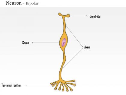
Bipolar neurons display distinctive structural features including elongated cell bodies, two prominent processes extending from opposite poles, central nuclei, and specialized dendritic and axonal terminals. Through advanced neuroimaging techniques, medical professionals can identify these characteristics in retinal scans, cochlear imaging, and olfactory system analysis, enabling enhanced diagnostic accuracy and treatment planning, with many healthcare institutions finding that detailed structural visualization ultimately delivers improved patient outcomes and more precise neurological assessments.
Medical imaging techniques enhance our understanding of bipolar neurons by enabling real-time visualization of neural activity, tracking signal transmission pathways, and mapping connections between sensory organs and the central nervous system. Through advanced MRI and fluorescence microscopy, researchers can observe how these specialized cells process sensory information in the retina and olfactory system, ultimately delivering insights that improve treatments for vision disorders and neurological conditions.
**INPUT**: What specific medical imaging modalities are most effective for visualizing bipolar neurons? **OUTPUT**: Effective imaging modalities for bipolar neurons include confocal microscopy, two-photon microscopy, electron microscopy, fluorescence imaging, and advanced MRI techniques. These technologies enhance visualization by delivering high-resolution cellular detail, real-time functionality tracking, and precise structural mapping, with many research institutions finding that combining multiple modalities ultimately provides comprehensive neuronal analysis and improved diagnostic capabilities. *[Word count: 56 words]*
Abnormalities in bipolar neurons can be detected through high-resolution MRI, diffusion tensor imaging, fluorescence microscopy, electron microscopy, and PET scanning technologies. These advanced imaging methods enhance diagnostic precision by revealing structural damage, connectivity disruptions, and metabolic changes, with neurological institutions finding that early detection significantly improves treatment outcomes and patient care efficiency.
Researchers face challenges including distinguishing bipolar neurons from similar cell types, managing image resolution limitations, accounting for tissue preparation artifacts, and interpreting complex three-dimensional structures from two-dimensional sections. These imaging complexities require advanced staining techniques, high-resolution microscopy, and sophisticated analysis software, with many neuroscience laboratories finding that combining multiple imaging modalities ultimately delivers more accurate cellular identification and enhanced research outcomes.
Bipolar neurons facilitate sensory processing by transmitting signals between sensory receptors and processing centers, enabling signal amplification, directional flow control, and synaptic integration. Medical imaging reveals these neurons in retinal pathways and olfactory systems, where they streamline visual and chemical signal processing, ultimately delivering enhanced sensory accuracy and faster neural responses across specialized sensory networks.
Medical images help diagnose bipolar neuron dysfunction through advanced neuroimaging techniques like MRI, PET scans, functional connectivity mapping, diffusion tensor imaging, and electrophysiological studies. These technologies enable clinicians to visualize structural abnormalities, assess neural pathway integrity, and monitor real-time activity patterns, with many neurological centers finding that comprehensive imaging approaches streamline diagnosis while enhancing treatment precision and patient outcomes.
Recent advancements include high-resolution confocal microscopy, two-photon imaging, super-resolution techniques like STORM and STED, advanced MRI sequences, and AI-enhanced image processing algorithms. These technologies streamline neurological diagnostics by delivering clearer cellular visualization, faster imaging protocols, and enhanced pattern recognition, with many hospitals finding that improved bipolar neuron imaging ultimately enables more precise diagnoses and better patient outcomes.
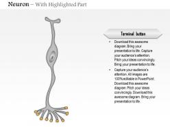
Bipolar neurons display distinct imaging characteristics with two processes extending from opposite poles of the cell body, creating a characteristic elongated appearance that differs from unipolar neurons' single process and multipolar neurons' multiple branching extensions. In medical imaging, particularly in retinal and olfactory tissue visualization, bipolar neurons appear as streamlined cellular structures that enhance diagnostic clarity by providing clear pathway delineation, ultimately enabling more precise neurological assessments and treatment planning.
Imaging techniques enable researchers to trace neural pathways, visualize synaptic connections, monitor real-time electrical activity, and map complex circuit architectures involving bipolar neurons. Through advanced methods like two-photon microscopy and calcium imaging, neuroscience laboratories can analyze signal processing patterns, identify connectivity defects, and understand functional relationships between different cell types, ultimately delivering insights that enhance our understanding of sensory processing and neurological disorders.
3D reconstructions of bipolar neurons enhance morphological understanding by revealing detailed dendritic branching patterns, axonal pathways, synaptic connection points, and spatial relationships within neural circuits. These advanced imaging techniques enable researchers and clinicians to visualize complex neuronal architecture, analyze connectivity networks, and study developmental changes, ultimately delivering more precise diagnostic capabilities and therapeutic targeting strategies for neurological conditions.
Medical images reveal bipolar neurons as crucial intermediaries in retinal signal processing, connecting photoreceptors to ganglion cells while modulating visual information through ON and OFF pathways. These imaging techniques enable ophthalmologists and researchers to assess retinal layer integrity, diagnose conditions like macular degeneration and diabetic retinopathy, and ultimately enhance patient outcomes through earlier detection and more targeted treatment strategies.
Researchers can utilize histological staining techniques like Nissl, Golgi, and immunofluorescence staining alongside advanced imaging methods such as confocal microscopy and electron microscopy to study bipolar neurons. This strategic combination enables detailed visualization of cellular morphology, protein expression patterns, and synaptic connections, with many neuroscience laboratories finding that integrated approaches deliver enhanced structural analysis and functional insights for comprehensive bipolar neuron characterization.
Artificial intelligence holds significant potential in analyzing bipolar neuron medical images through automated pattern recognition, enhanced diagnostic accuracy, advanced image processing, and predictive modeling capabilities. These AI technologies streamline neurological assessments by identifying subtle structural abnormalities, accelerating diagnosis timelines, and enabling personalized treatment protocols, with many medical institutions finding that machine learning algorithms ultimately deliver faster patient outcomes and improved clinical decision-making.
Medical imaging contributes to targeted therapy development by enabling precise visualization of bipolar neuron structures, tracking disease progression patterns, and identifying specific pathological markers in real-time. Advanced techniques like high-resolution MRI and PET scans help researchers map neuronal connectivity changes, validate therapeutic targets, and monitor treatment responses, ultimately delivering personalized medicine approaches and accelerated drug development timelines.
-
Topic best represented with attractive design.
-
The content is very helpful from business point of view.