

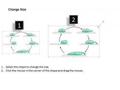
0614 steps in cell motility medical images for powerpoint





Try Before you Buy Download Free Sample Product
 Impress Your
Audience
 100%
Editable
 Save Hours
of Time


Enhance the ecosystem with our 0614 Steps In Cell Motility Medical Images For Powerpoint. Enlist yourself in the cause of conservation.
People who downloaded this PowerPoint presentation also viewed the following :
0614 steps in cell motility medical images for powerpoint with all 10 slides:
Accessorise your thoughts with our 0614 Steps In Cell Motility Medical Images For Powerpoint. Give them the extra glow they deserve.




FAQs for 0614 steps in cell motility medical
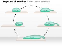
There are basically three ways cells move around. Actin polymerization is what most crawling cells use - like fibroblasts pushing out those lamellipodia things at the front. Flagella and cilia work more like whips, which is how sperm swim (and some bacteria too). Myosin contraction is the squeezing mechanism - you see this in amoebas and immune cells that have to squeeze through tight spots. Real cells are messier though. They usually mix these mechanisms depending on what they're doing. If you're planning experiments, figure out which one your cell type relies on most. That'll save you time.
So ECM proteins are like roadways for cells - they literally grab onto fibronectin and laminin through integrins to move around. Collagen gives them tracks to follow too. Here's the weird part: stiffness totally matters. Cells hate surfaces that are super squishy or rock hard, but they love that middle ground (classic Goldilocks situation). You can actually steer cells around using ECM gradients since they'll follow concentration trails. Oh, and if you're setting up migration assays, definitely try coating with different ECM proteins - you'll see crazy differences in how your cells behave.
So actin and microtubules are like the cell's whole movement system. Actin filaments do the actual crawling - they build up at the front edge and literally push the membrane forward. Pretty cool mechanism honestly. Microtubules are more like the transport highways, moving stuff around and keeping everything organized. You've also got actin forming these contractile bundles that yank the cell's back end forward. The coordination between both systems is what makes migration work. If you mess with either one using drugs like cytochalasin D or nocodazole, the whole thing falls apart.
So basically, single-celled organisms are like little swimmers - their whole body moves using flagella or those stretchy pseudopodia things. Pretty straightforward. But with multicellular organisms? Most cells stay put (which is good because otherwise we'd literally fall apart lol), except for specific ones like sperm or immune cells that can actually migrate through tissues. The cool part is they use similar molecular machinery, but the reasons are totally different. Single cells are just trying to find food or avoid getting eaten, while our cells move for stuff like development or immune responses. Honestly, if you're studying this, focus on comparing those underlying mechanisms - that's where it gets interesting.
So basically cells follow chemical signals - growth factors, cytokines, stuff like that. They bind to receptors and kick off these cascades. What's cool is how the cell sets up this polarity thing: PI3K/Akt gets active at the front while PTEN hangs out at the back. Creates a nice gradient. Rho GTPases are super important too - Rac1 pushes the front forward, RhoA pulls the back. Calcium also tweaks how sensitive everything is. If you're gonna test this out, I'd start simple with EGF or PDGF gradients. Way easier than jumping into the fancy stuff right away.
So basically, cancer spreads through metastasis, right? Cell motility research helps us figure out exactly how that happens. Normal cells move and stick to tissues in predictable ways, but cancer cells? They're total rebels - their cytoskeleton gets all messed up and they become invasive. Scientists are using this info to create drugs that block specific movement pathways. Maybe they can stop metastasis before it even starts. There's some cool stuff happening with integrin inhibitors and Rho kinase blockers in clinical trials right now if you're curious.
So there's a few main ways to do this. Live fluorescence microscopy is probably your best bet - you can track specific proteins or just label the whole cell with GFP. Phase contrast works well too if you don't want to deal with labeling, plus it shows morphology changes really clearly. Traction force microscopy is cool if you need to measure actual forces (uses deformable substrates). Honestly watching cells migrate in real-time never gets old, it's weirdly addictive. For fast stuff you might need confocal systems but that's getting fancy. I'd probably just start with basic phase contrast though.
Dude, cell motility is literally how wounds heal. Your immune cells have to migrate over to clean up all the junk and fight off bacteria. Fibroblasts crawl in next to build new tissue - kinda like construction workers. Then epithelial cells actually walk across the wound to seal it shut, which is honestly pretty crazy if you picture it happening. That's why diabetics have such a hard time healing - high blood sugar screws with how cells move around. Oh, and if you're researching this stuff, definitely look at the motility pathways first since that's where things usually go wrong.
Yeah so basically when cells can't move around properly, they just pile up in one spot and start cranking out tons of collagen. Think of it like workers stuck in one corner of a construction site - they just keep building there instead of spreading out. That's how you get those rigid, scarred areas in lung fibrosis and liver disease. The fibroblasts go crazy with ECM production since they can't migrate where they're supposed to. Pretty fascinating stuff actually - you should definitely look into mechanotransduction pathways if you're diving deeper into this.
Think of motor proteins as the cell's moving crew and construction workers rolled into one. They crawl along microtubules and actin filaments to totally reshape the cytoskeleton. This lets cells do crazy stuff - squeezing through tiny gaps, climbing over barriers, pushing through resistance. Myosin's the MVP here, contracting the back end while other motors stretch out the front. Creates this coordinated crawling thing you see when immune cells hunt down infections (honestly pretty wild to watch under a microscope). Don't just study each motor type separately though - it's all about how they work together.
Yeah so basically immune cells have to move around or they're useless. Like when you get an infection, neutrophils and macrophages follow chemical signals to track down the bad stuff and eat it. T cells and B cells do the same thing but in your lymph nodes to find antigens. The whole system falls apart without motility - imagine security guards stuck at their desk while burglars run around the building lol. Pretty wild how they navigate through all that tissue mess honestly. If you're looking at immune problems, motility issues are definitely worth checking out.
Oh man, the bioengineering stuff for cell migration is seriously cool. Microfluidics lets you create these super precise chemical gradients - basically steering cells wherever you want. Then there's optogenetics where you can literally use light to flip proteins on and off in real time. I spent way too much time last week just watching cells respond under the microscope, it's oddly mesmerizing. You can totally reprogram how cells move now, which would've been impossible a few years ago. If you're diving into migration work, definitely look into the new optogenetic actuators - they're incredible for messing with cytoskeletal dynamics.
So basically, cells have two ways to move around obstacles. Amoeboid cells just squeeze through tight spots by changing their shape - super fast but only works if the gap's big enough. Mesenchymal cells are totally different - they actually break down whatever's in their way by pumping out enzymes. Way slower but they can get through anything. Cancer cells are honestly pretty clever because they'll switch between both methods depending on what they hit. If you're looking at this stuff in the lab, check for MMPs to spot mesenchymal movement, or just see if your cells can fit through tiny spaces for amoeboid behavior.
So basically, mechanical forces are like traffic cops for your cells - they control where and how fast cells migrate. Stretch forces pull cells in that direction, while compression usually slows them down or makes them go elsewhere. Cells actually "sense" these mechanical cues, which is pretty crazy if you think about it. Stiffer surfaces make cells move faster and more directionally too. Oh, and if you're doing any migration experiments, the mechanical setup you use will totally change your results compared to real tissue - learned that one the hard way!
So basically, these bioactive compounds mess with how cells move around by targeting the pathways that control their "skeleton" and migration stuff. Cytochalasins will wreck actin filaments, while other compounds go after Rho GTPases or integrin signaling - they're literally hijacking the cell's movement controls. Growth factors make cells move more, but some compounds completely freeze them. It's honestly crazy how one tiny molecule can make cells either sprint everywhere or just stop dead. When you're looking at this stuff, figure out which pathway gets hit first - that's what'll tell you if you're getting more or less cell movement.
-
I discovered this website through a google search, the services matched my needs perfectly and the pricing was very reasonable. I was thrilled with the product and the customer service. I will definitely use their slides again for my presentations and recommend them to other colleagues.
-
Editable templates with innovative design and color combination.