

0714 axillary artery medical images for powerpoint





Try Before you Buy Download Free Sample Product
 Impress Your
Audience
 100%
Editable
 Save Hours
of Time


Bring your team on the same page as you. Our 0714 Axillary Artery Medical Images For PowerPoint will put them in sync.
People who downloaded this PowerPoint presentation also viewed the following :
0714 axillary artery medical images for powerpoint with all 10 slides:
Pitching the perfect project can be stressful. With our 0714 Axillary Artery Medical Images For PowerPoint we take care of the design for you.







FAQs for 0714 axillary artery medical
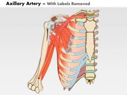
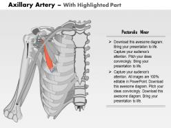
The axillary artery comprises three anatomical divisions based on its relationship to the pectoralis minor muscle: first part (medial to the muscle), second part (posterior to the muscle), and third part (lateral to the muscle). These divisions systematically organize the artery's branching pattern and surgical approaches, with medical professionals finding that this classification enhances procedural precision and educational understanding.
The axillary artery extends from the first rib to the lower border of the teres major muscle, while the brachial artery continues from that point down the upper arm to the elbow. The axillary artery primarily supplies the shoulder region, chest wall, and axillary structures, whereas the brachial artery delivers blood to the arm muscles and forearm, with the axillary serving as the major transitional vessel between central circulation and peripheral arm blood supply.
The axillary artery gives rise to six major branches including the superior thoracic, thoracoacromial, lateral thoracic, subscapular, anterior circumflex humeral, and posterior circumflex humeral arteries. These vessels supply the shoulder girdle, chest wall, and upper arm muscles, with the circumflex branches forming crucial collateral circulation around the shoulder joint, ensuring continuous blood flow during arm movement and potential vascular compromise.
The axillary artery delivers oxygenated blood to the upper limb through six major branches, including the superior thoracic, thoracoacromial, lateral thoracic, subscapular, and anterior/posterior circumflex humeral arteries. These vessels systematically supply the shoulder muscles, joint capsule, and surrounding tissues, ensuring comprehensive circulation that supports arm mobility, muscle function, and tissue repair throughout the shoulder girdle and proximal arm region.
Axillary artery injuries present significant clinical challenges including potential limb ischemia, compartment syndrome, nerve damage, and vascular compromise requiring immediate surgical intervention. These injuries, commonly seen in trauma centers and orthopedic practices, demand rapid assessment and repair to prevent permanent disability, with many vascular surgeons finding that early recognition and treatment ultimately deliver better functional outcomes and limb preservation.
The axillary artery runs alongside the brachial plexus through the axilla, with both structures intimately related as the artery transitions from subclavian to brachial artery. This anatomical relationship is crucial for surgeons and clinicians, as procedures involving axillary lymph node dissection or shoulder surgeries require careful navigation to avoid injury to both vascular and neural structures, ultimately ensuring optimal patient outcomes.
The axillary artery serves as a critical access point for vascular surgeries, bypass procedures, and catheter-based interventions targeting upper limb circulation. Through its strategic positioning and robust collateral network, surgeons can perform arterial reconstructions, treat occlusive diseases, and establish vascular access, while its anatomical landmarks enable precise surgical planning, ultimately delivering improved limb perfusion and functional outcomes.
The axillary artery can become compressed in thoracic outlet syndrome when anatomical structures like ribs, muscles, or scar tissue narrow the thoracic outlet space, reducing blood flow to the arm. This compression manifests through symptoms including arm pain, numbness, weakness, and cold sensitivity, with severe cases potentially leading to blood clots or aneurysm formation requiring surgical intervention.
Ultrasound, CT angiography, MR angiography, digital subtraction angiography, and duplex scanning are most effective for visualizing the axillary artery. These imaging modalities enhance diagnostic accuracy by providing detailed vascular mapping, real-time blood flow assessment, and comprehensive anatomical visualization, with many medical institutions finding that combining multiple techniques delivers superior surgical planning and patient outcomes.
The axillary artery's position shifts dynamically as the arm moves, with abduction elevating and stretching the vessel while adduction relaxes its positioning against surrounding muscular structures. This anatomical flexibility enables consistent blood flow during various arm positions, with surgeons and interventional specialists finding that understanding these positional changes enhances procedural accuracy and reduces vascular complications during shoulder procedures.
Risks include bleeding, hematoma formation, nerve injury, thrombosis, and vessel dissection or perforation. These complications can lead to limb ischemia, brachial plexus damage, or infection, with many cardiovascular centers finding that careful patient selection, advanced imaging guidance, and experienced surgical teams significantly minimize these risks while delivering improved outcomes for complex vascular procedures.
During physical activity, blood flow in the axillary artery increases significantly through vasodilation, enhanced cardiac output, and reduced peripheral resistance to meet elevated metabolic demands of arm and shoulder muscles. This physiological adaptation enables improved oxygen delivery, nutrient transport, and waste removal, while maintaining optimal circulation patterns that support sustained muscular performance and efficient recovery processes.
Diagnostic procedures for axillary artery assessment include Doppler ultrasound, CT angiography, MR angiography, digital subtraction angiography, and arterial pressure measurements. These imaging technologies enhance diagnostic accuracy by visualizing vessel anatomy, detecting stenosis or occlusion, and measuring blood flow patterns, with many vascular laboratories finding that combining multiple modalities delivers comprehensive evaluation and optimal treatment planning.
The axillary artery engages robust collateral circulation through interconnected networks including the thoracoacromial, lateral thoracic, subscapular, and circumflex humeral branches, which create alternative pathways around potential blockages. These vascular networks enable continued blood flow to the upper extremity by establishing connections with intercostal arteries and scapular vessels, ultimately maintaining tissue perfusion and functional capacity even when primary arterial flow is compromised.
The axillary artery serves as a critical alternative pathway for cardiac circulation when coronary arteries become compromised by disease, enabling surgeons to perform bypass grafts and revascularization procedures. In ischemic conditions, this vessel provides essential collateral blood flow to heart muscle, with cardiac surgeons increasingly utilizing axillary access for minimally invasive procedures, ultimately delivering improved patient outcomes and reduced surgical complications.
-
Great designs, really helpful.
-
Good research work and creative work done on every template.