

0714 dynamic instability of microtubules medical images for powerpoint





Try Before you Buy Download Free Sample Product
 Impress Your
Audience
 100%
Editable
 Save Hours
of Time


Point the needle with our 0714 Dynamic Instability Of Microtubules Medical Images For Powerpoint. Direct your thoughts to the path they need to take.
People who downloaded this PowerPoint presentation also viewed the following :
0714 dynamic instability of microtubules medical images for powerpoint with all 9 slides:
Our 0714 Dynamic Instability Of Microtubules Medical Images For Powerpoint bring it close at hand. Achieving desired goals becomes a certainty.




FAQs for 0714 dynamic instability of microtubules medical
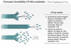
Microtubules consist of α- and β-tubulin heterodimers that polymerize into protofilaments, with GTP-binding sites enabling structural transitions between stable and unstable states. These protein subunits create dynamic assembly and disassembly cycles through GTP hydrolysis, allowing cellular structures to rapidly reorganize during mitosis, intracellular transport, and cytoskeletal remodeling processes.
The GTP cap creates a stabilizing buffer zone at growing microtubule ends by maintaining structural integrity through GTP-bound tubulin subunits, preventing catastrophic depolymerization events. This mechanism enables cells to regulate cytoskeletal dynamics precisely, with many organizations finding that understanding microtubule stability enhances drug development strategies, particularly in cancer therapeutics and neurological research applications.
**INPUT**: What role does the highly regulated cycle of tubulin GTP hydrolysis play in microtubule dynamics? **OUTPUT**: The highly regulated cycle of tubulin GTP hydrolysis serves as the fundamental molecular switch controlling microtubule assembly and disassembly, creating the essential dynamic instability that enables cellular reorganization. This strategic mechanism delivers rapid cytoskeletal restructuring, precise chromosome segregation, and adaptive cellular responses, ultimately enabling cells to efficiently navigate division, migration, and intracellular transport processes.
Temperature and cellular conditions significantly influence microtubule dynamic instability by altering polymerization rates, catastrophe frequency, and rescue events through changes in tubulin concentration, GTP hydrolysis kinetics, and regulatory protein activity. Higher temperatures generally accelerate both assembly and disassembly processes, while cellular factors like microtubule-associated proteins, calcium levels, and ATP availability modulate stability, with many research applications finding that controlled conditions enable precise cytoskeletal manipulation.
Microtubule dynamic instability enables mitotic spindle formation by allowing rapid assembly and disassembly of spindle fibers, precise chromosome capture through search-and-capture mechanisms, and accurate segregation during anaphase. This controlled instability facilitates efficient cell division by enabling spindle checkpoint responses, ensuring proper chromosome alignment, and allowing timely spindle disassembly, ultimately delivering reliable cellular reproduction essential for tissue growth and organismal development.
Microtubule-associated proteins (MAPs) regulate microtubule stability through stabilization, destabilization, and dynamic modulation mechanisms. These regulatory proteins enhance cellular processes by controlling spindle formation during mitosis, facilitating intracellular transport pathways, and enabling rapid cytoskeletal reorganization, with research institutions finding that MAP dysfunction significantly impacts neurological development and cancer progression.
Live-cell microtubule dynamics are studied using fluorescence microscopy techniques including live imaging with fluorescently-labeled tubulin, photobleaching methods like FRAP and photoactivation, and advanced approaches such as speckle microscopy and plus-end tracking proteins. These methodologies enable researchers to quantify growth rates, catastrophe frequencies, and rescue events in real-time, with many cell biology laboratories finding that combining multiple techniques delivers comprehensive insights into microtubule behavior across different cellular contexts.
Post-translational modifications of tubulin significantly influence microtubule stability by regulating polymerization rates, depolymerization patterns, and motor protein interactions through acetylation, detyrosination, and phosphorylation. These modifications enable cells to fine-tune cytoskeletal dynamics for specific functions like mitotic spindle formation, axonal transport in neurons, and cellular migration, ultimately delivering enhanced organizational control and adaptive responses.
Therapeutic strategies targeting microtubule dynamics include taxanes like paclitaxel, vinca alkaloids, eribulin, and novel agents like KSP inhibitors and aurora kinase inhibitors. These treatments disrupt cancer cell division by stabilizing or destabilizing microtubules, with pharmaceutical companies increasingly developing combination therapies that enhance efficacy while minimizing resistance, ultimately delivering improved patient outcomes across multiple cancer types.
Microtubules enable cellular motility and transport by providing dynamic tracks for motor proteins like dynein and kinesin, facilitating organelle movement, vesicle trafficking, and chromosome segregation during mitosis. Their dynamic instability allows rapid reorganization of transport networks, enabling cells to quickly redirect cargo flows, establish new migration pathways, and adapt cytoskeletal architecture for efficient intracellular logistics and coordinated cellular movement.
Dynamic instability enables microtubules to rapidly reorganize cellular architecture during neurogenesis and synaptic plasticity by alternating between growth and shrinkage phases, facilitating axon guidance, dendritic branching, and synaptic remodeling. This controlled flexibility supports neural development, memory formation, and adaptive responses, with neurons leveraging these structural changes to establish new connections, strengthen existing pathways, and ultimately enhance learning capacity and neuroplasticity.
Microtubules exhibit catastrophic depolymerization and rescue events, while actin filaments undergo more gradual treadmilling with steady polymerization and depolymerization at opposite ends. This difference enables microtubules to rapidly reorganize cellular architecture during mitosis and intracellular transport, whereas actin provides more stable structural support for cell motility, with many cell biology researchers finding that this combination delivers both flexibility and structural integrity for complex cellular functions.
Mechanical forces significantly influence microtubule stability through tension, compression, and cellular stresses that can either stabilize or destabilize these structures during mitosis, migration, and organelle transport. These forces enable cells to dynamically reorganize their cytoskeleton in response to environmental changes, with muscle cells, neurons, and dividing cells demonstrating enhanced structural adaptation and coordinated cellular functions under varying mechanical conditions.
Understanding microtubule dynamics is crucial for neurodegenerative disease research because disrupted microtubule stability directly contributes to neuronal dysfunction, protein aggregation, and cellular transport failures. This knowledge enables pharmaceutical companies and research institutions to develop targeted therapies that restore microtubule function, ultimately delivering improved treatment outcomes for conditions like Alzheimer's and Parkinson's disease.
Computational modeling enables prediction of microtubule behavior by simulating polymerization dynamics, catastrophe frequencies, and rescue events under varying ATP concentrations, temperature fluctuations, and protein interactions. These models help researchers in pharmaceutical companies, biotechnology firms, and academic institutions optimize drug design targeting cancer cells, predict cellular responses to treatments, and accelerate therapeutic development timelines.
-
Professional and unique presentations.
-
Illustrative design with editable content. Exceptional value for money. Highly pleased with the product.