

0814 ovarian cancer medical images for powerpoint





Try Before you Buy Download Free Sample Product
 Impress Your
Audience
 100%
Editable
 Save Hours
of Time


Our 0814 Ovarian Cancer Medical Images For Powerpoint have a refreshing effect. They will reactivate the energies of your team.
People who downloaded this PowerPoint presentation also viewed the following :
0814 ovarian cancer medical images for powerpoint with all 10 slides:
Keep them glued with our 0814 Ovarian Cancer Medical Images For Powerpoint. They may even forget to blink.

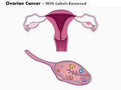
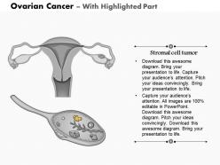
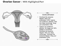


FAQs for 0814 ovarian cancer medical
The most common imaging techniques for ovarian cancer diagnosis include transvaginal ultrasound, CT scans, MRI, PET scans, and contrast-enhanced imaging protocols. These technologies enhance diagnostic accuracy by providing detailed tumor visualization, assessing metastatic spread, and enabling precise staging, with many oncology centers finding that strategic combination of multiple modalities ultimately delivers faster treatment planning and improved patient outcomes.
MRI excels at soft tissue contrast and characterizing complex ovarian masses, while CT scans provide faster imaging and superior detection of calcifications, metastases, and overall staging. Both modalities complement each other in clinical practice, with many oncologists finding that MRI's detailed pelvic anatomy visualization combined with CT's rapid assessment capabilities delivers comprehensive diagnostic accuracy, ultimately enabling more precise treatment planning and improved patient outcomes.
Ultrasound serves as a primary screening tool for ovarian cancer detection through transvaginal imaging that identifies ovarian masses, cysts, and structural abnormalities with enhanced clarity and precision. While particularly effective when combined with blood markers like CA-125, ultrasound enables gynecologists and oncologists to detect suspicious changes early, ultimately delivering faster diagnostic pathways and improved treatment outcomes for patients.
Yes, several imaging biomarkers significantly enhance ovarian cancer diagnosis, including papillary projections, solid components, septations, ascites, and vascular patterns on ultrasound and MRI. These biomarkers enable radiologists to differentiate malignant from benign masses more accurately, with many oncology centers finding that combining multiple biomarkers improves diagnostic precision and ultimately delivers faster treatment decisions.
Ovarian cancer staging is accurately assessed through CT scans, MRI, PET scans, and ultrasound imaging, which evaluate tumor size, lymph node involvement, and metastatic spread. These imaging technologies streamline diagnostic processes by detecting peritoneal implants, ascites, and distant metastases, enabling oncologists to develop precise treatment protocols and ultimately delivering improved patient outcomes and more targeted therapeutic approaches.
Interpreting ovarian cancer medical images presents challenges including subtle early-stage lesions, overlapping benign conditions, complex pelvic anatomy, variable tumor appearances, and imaging artifact interference. These diagnostic complexities are increasingly addressed through advanced AI-powered imaging analysis and specialized radiological training, with many healthcare institutions finding that enhanced imaging protocols and machine learning integration significantly improve diagnostic accuracy and treatment outcomes.
Imaging technologies help differentiate ovarian masses through advanced MRI sequences, contrast-enhanced CT scans, ultrasound elastography, and PET imaging that analyze tissue characteristics, vascular patterns, and metabolic activity. These diagnostic tools enable radiologists to identify specific markers like restricted diffusion, enhancement patterns, and morphological features, ultimately delivering more accurate diagnoses and helping oncologists develop targeted treatment strategies for improved patient outcomes.
**INPUT**: What advancements in imaging technology have the potential to enhance ovarian cancer screening? **OUTPUT**: Emerging imaging advancements include AI-enhanced ultrasound, molecular imaging with targeted contrast agents, multiparametric MRI, photoacoustic imaging, and advanced CT techniques with texture analysis. These technologies streamline early detection by improving tissue characterization, reducing false positives, and enabling automated analysis, with many healthcare institutions finding that strategic integration delivers faster diagnostic workflows and enhanced patient outcomes. [Word count: 58 words]
Contrast-enhanced imaging improves ovarian tumor visualization by increasing tissue differentiation, enhancing vascular patterns, and highlighting tumor boundaries against surrounding healthy tissue. Through advanced contrast agents, radiologists can detect smaller lesions, assess tumor vascularity, and differentiate between benign and malignant masses more accurately, ultimately enabling earlier detection and more precise treatment planning.
3D imaging revolutionizes ovarian cancer surgical planning by providing detailed anatomical visualization, precise tumor mapping, enhanced depth perception, and improved spatial relationships between organs and malignancies. Through advanced reconstruction technologies, surgeons can simulate procedures, identify optimal approaches, and minimize complications, while ultimately delivering more precise resections and better patient outcomes in an increasingly complex surgical landscape.
PET scans evaluate ovarian cancer treatment response by measuring metabolic activity changes in tumor tissues, detecting residual disease, and identifying new metastases through glucose uptake patterns. These imaging capabilities enable oncologists to assess chemotherapy effectiveness, modify treatment protocols when necessary, and monitor patient progress more precisely, ultimately delivering personalized care strategies and improved therapeutic outcomes.
Imaging findings significantly influence ovarian cancer prognosis by revealing tumor stage, peritoneal spread, lymph node involvement, and response to treatment, directly impacting survival rates and therapeutic decisions. Advanced imaging enables oncologists to stratify patients into risk categories, optimize surgical planning, and monitor treatment effectiveness, with many cancer centers finding that comprehensive imaging assessment ultimately delivers more personalized care and improved patient outcomes.
Imaging findings vary significantly by histology, with serous carcinomas typically showing complex cystic masses with solid components and peritoneal implants, while mucinous tumors present as large multilocular cystic lesions with variable enhancement patterns. These histologic differences enable radiologists to provide more targeted diagnostic insights, ultimately enhancing treatment planning accuracy and improving patient outcomes across oncology departments.
Radiologists serve as critical diagnostic partners in ovarian cancer management, providing expertise in initial detection, staging accuracy, treatment monitoring, and recurrence surveillance through advanced imaging techniques. Working closely with gynecologic oncologists, pathologists, and medical oncologists, they guide surgical planning, assess chemotherapy responses, and detect disease progression, ultimately enabling personalized treatment strategies and improved patient outcomes across comprehensive cancer care teams.
Artificial intelligence enhances ovarian cancer imaging through automated pattern recognition, advanced image analysis algorithms, and predictive modeling systems that identify subtle abnormalities in CT scans, MRIs, and ultrasounds. These AI-powered tools streamline radiologist workflows, improve diagnostic accuracy, and enable earlier detection, with many healthcare institutions finding that integrated AI systems ultimately deliver faster diagnosis times and more precise treatment planning.
-
Awesome presentation, really professional and easy to edit.
-
Presentation Design is very nice, good work with the content as well.