

Pituitary Tumor Surgery Craniotomy PPT Template ACP





Try Before you Buy Download Free Sample Product
 Impress Your
Audience
 100%
Editable
 Save Hours
of Time


Elevate your presentations with our Pituitary Tumor Surgery Craniotomy PPT Template. This professional deck features comprehensive slides, detailed visuals, and expert insights, designed for medical professionals. Effectively communicate complex concepts, enhance understanding, and engage your audience in discussions about pituitary tumor surgical techniques and outcomes. Perfect for conferences and educational settings.
People who downloaded this PowerPoint presentation also viewed the following :
Pituitary Tumor Surgery Craniotomy PPT Template ACP with all 41 slides:
Use our Pituitary Tumor Surgery Craniotomy PPT Template ACP to effectively help you save your valuable time. They are readymade to fit into any presentation structure.
























FAQs for Pituitary Tumor Surgery Craniotomy
Pituitary tumors include functioning adenomas (prolactinomas, growth hormone-secreting, ACTH-producing), non-functioning adenomas, craniopharyngiomas, and Rathke's cleft cysts, each requiring distinct treatment protocols. These conditions vary significantly in surgical approach, with endoscopic transsphenoidal surgery preferred for most adenomas, while larger tumors may require transcranial approaches, ultimately delivering targeted treatment plans that restore hormonal balance and preserve neurological function.
Common symptoms indicating surgical intervention include severe headaches, vision problems like peripheral vision loss, hormonal imbalances causing fatigue or reproductive issues, unexplained weight changes, and cognitive difficulties. These symptoms often worsen progressively, with many patients finding that surgical removal through minimally invasive techniques significantly restores quality of life, hormonal balance, and visual function while preventing further neurological complications.
The decision for pituitary tumor surgery depends on tumor size, hormone production levels, symptom severity, and patient health status. Neurosurgeons and endocrinologists collaborate to evaluate factors like visual field defects, hormonal imbalances, and growth patterns, with many medical centers finding that comprehensive imaging and biochemical testing ultimately delivers personalized treatment plans and optimal patient outcomes.
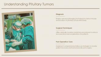
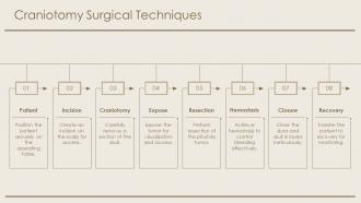
Common pituitary tumor surgical techniques include transsphenoidal endoscopic surgery, microscopic transsphenoidal approach, transcranial surgery, and extended endonasal procedures. These approaches enhance patient outcomes by minimizing invasive access, reducing recovery times, and preserving critical neurological functions, with many medical centers finding that endoscopic techniques deliver improved visualization and precision.
Endoscopic surgery serves as the preferred minimally invasive approach for pituitary tumor removal, offering enhanced visualization, precise tumor resection, and reduced surgical trauma compared to traditional methods. Through advanced endoscopic techniques, neurosurgeons achieve improved access to complex anatomical areas, faster patient recovery, and lower complication rates, with many medical centers finding that this approach delivers superior outcomes while minimizing hospital stays and enhancing overall treatment effectiveness.
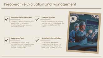
Pre-operative MRI significantly influences surgical planning by revealing tumor size, location, carotid artery proximity, optic chiasm involvement, and sphenoid sinus anatomy. These detailed imaging insights enable neurosurgeons to determine optimal surgical approaches, anticipate potential complications, and customize instrumentation selection, ultimately delivering safer procedures, reduced operative times, and improved patient outcomes across specialized medical centers.
Potential risks include cerebrospinal fluid leaks, hormone deficiencies, vision changes, infection, and bleeding complications. While most patients experience successful outcomes with minimal complications, some may require hormone replacement therapy or additional treatments, with many medical centers finding that experienced neurosurgical teams significantly reduce these risks through advanced techniques.
Patients can prepare for pituitary tumor surgery through comprehensive medical evaluation, optimizing overall health, discussing concerns with their surgical team, and establishing realistic recovery expectations. Many patients find that maintaining physical fitness, arranging post-operative support systems, and understanding the surgical process reduces anxiety while enhancing recovery outcomes, ultimately delivering better surgical experiences and faster healing.
**INPUT**: What factors determine the likelihood of a successful outcome following pituitary tumor surgery? **OUTPUT**: Successful pituitary tumor surgery outcomes depend on tumor size, location, type, patient age, overall health status, and surgeon expertise with specialized techniques. These factors work together to influence recovery rates, hormone function restoration, and symptom resolution, with many medical centers finding that early detection and minimally invasive approaches significantly enhance patient outcomes and long-term neurological function. [Word count: 57 words]
Post-operative care for pituitary tumor surgery includes hormone level monitoring, nasal packing management, fluid balance assessment, neurological evaluations, and gradual activity resumption. Through specialized endocrine protocols, medical teams streamline recovery by preventing complications like diabetes insipidus, managing potential hormone deficiencies, and ensuring optimal healing, with many patients finding that comprehensive follow-up care ultimately delivers faster recovery and better long-term outcomes.
Hormonal imbalances from pituitary tumor surgery are managed through hormone replacement therapy, regular monitoring, and multidisciplinary care coordination involving endocrinologists and neurosurgeons. Treatment protocols include thyroid hormone supplements, cortisol replacement, and growth hormone therapy when indicated, with many medical centers finding that personalized dosing schedules and frequent laboratory assessments enable optimal patient outcomes and improved quality of life.
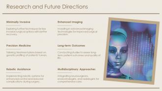
Advancements include endoscopic endonasal surgery, intraoperative MRI, neuronavigation systems, fluorescence-guided surgery, and enhanced imaging technologies. These innovations streamline surgical precision by minimizing tissue damage, improving tumor visualization, and enabling real-time guidance, with many neurosurgical centers finding that patients experience faster recovery times, reduced complications, and enhanced preservation of pituitary function.
A multi-disciplinary approach integrates neurosurgeons, endocrinologists, radiologists, pathologists, and rehabilitation specialists to optimize patient outcomes through comprehensive care coordination, precise hormone management, and tailored recovery protocols. This collaborative model enhances surgical precision, accelerates post-operative recovery, and minimizes complications, with many medical centers finding that coordinated specialist teams deliver superior long-term patient outcomes and faster functional restoration.
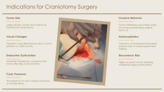
Tumor size and location directly determine whether neurosurgeons use minimally invasive transsphenoidal approaches through the nasal cavity or more extensive craniotomy procedures, with smaller pituitary adenomas typically managed endoscopically. Large tumors extending beyond the sella turcica or those compressing critical structures like the optic chiasm require strategic surgical planning, ultimately enabling surgeons to minimize complications while maximizing tumor removal and preserving neurological function.
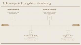
Post-surgical monitoring protocols include regular MRI scans every 6-12 months initially, comprehensive hormonal assessments to evaluate pituitary function, visual field testing, and clinical examinations. These systematic approaches enable early detection of tumor recurrence, assessment of surgical success, and identification of hormonal deficiencies, with many medical centers finding that structured follow-up protocols ultimately deliver improved patient outcomes and enhanced long-term management strategies.
-
“There is so much choice. At first, it seems like there isn't but you have to just keep looking, there are endless amounts to explore.”
-
“Immediate response, professional support, and effective solutions that were customized and immediately provided. Well done- Thank you!”