


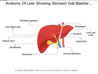
Anatomy of liver showing stomach gall bladder light right lobe




Try Before you Buy Download Free Sample Product
 Impress Your
 Audience
 100%
100% Editable
 Save Hours
 of Time


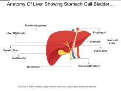
Be able to focus your energies again with our anatomy of liver showing stomach gall bladder light right lobe PPT deck and bring back that clear locus. Through the example of this PowerPoint design, we have shown the working of liver and gall balder. small components like a falciform ligament, liver light robe, hepatic duct, gall bladder, common bile duct, cystic duct, stomach, liver left lobe and esophagus are shown in this PPT layout to give a piece of complete information on the components of the human liver. Take your process to the next stop with our, anatomy of liver showing stomach gall bladder light right lobe PPt deck and download this slide to get ahead. Be able to concentrate your energies again with our Anatomy Of Liver Showing Stomach Gall Bladder Light Right Lobe. Bring back that clear focus.
People who downloaded this PowerPoint presentation also viewed the following :
Anatomy of liver showing stomach gall bladder light right lobe with all 5 slides:
Extract the absolute best with our Anatomy Of Liver Showing Stomach Gall Bladder Light Right Lobe. They enable you to drive a hard bargain.


FAQs for Anatomy of liver showing stomach gall bladder
So the liver's got two main parts - right and left lobes split by this thing called the falciform ligament. Right side is massive, like 6x bigger than the left. Then there's two tiny lobes hiding in back: the quadrate lobe (squeezed between gallbladder and ligamentum teres) and caudate lobe behind the left near the IVC. Honestly though, if you're doing surgery stuff, forget all that anatomy textbook nonsense. Couinaud's eight segments make way more sense since they actually follow how blood flows and bile drains. Way more practical.
So the liver's blood supply is actually pretty wild - it gets fed by two different sources instead of just one like most organs. There's the hepatic artery bringing in fresh oxygenated blood, but then it also gets this nutrient-packed blood from the portal vein that's already made a pit stop at your intestines. About 75% comes from that portal vein, 25% from the artery. It's honestly such a smart design when you think about it. But yeah, this dual setup means if either system gets messed up during surgery or disease, you're gonna have problems.
So the portal triad is basically three structures bundled together in liver tissue - you've got the hepatic artery bringing oxygen-rich blood, portal vein carrying nutrients from your gut, and a bile duct for drainage. It's like the liver's pit stop honestly. Blood flows from these areas toward the central vein in each lobule. When I was studying histology slides, spotting the triad first made everything else click into place - you can orient yourself and figure out which way blood's moving. Pretty useful for understanding how hepatocytes get fed and dump their waste.
So liver lobules are these hexagonal little units that do all the actual work. Picture tiny factories with a central vein in the middle and liver cells spreading out like wheel spokes. Portal triads hang out at the corners - they've got bile ducts, arteries, and veins all bundled together. Blood flows inward while bile goes the opposite way, which is honestly pretty clever design. Oh and here's the thing - if you're studying liver diseases later, this whole setup is key. Diseases follow these patterns, so once you get the basic architecture down, pathology starts making way more sense.
Your gallbladder sits in this little depression on the bottom of your liver - right between the right and quadrate lobes. Think of it as roommates sharing the same apartment. Blood comes from the cystic artery (usually branches off the right hepatic), and drainage goes straight back into the liver tissue or portal system. Here's the thing though - because they're so close together, when one gets sick, the other usually does too. That's why radiologists always look at both on scans. Actually makes sense when you think about the anatomy.
So the liver's basically wedged under your diaphragm covering both upper quadrants, which totally changes how you approach it. Most surgeries go subcostal or upper midline because you can't really get at it from above - heart and lungs are in the way. Major vessels like the IVC run right next to it, so you need solid visualization before you start cutting. Honestly, the posterior segments are a pain laparoscopically since everything's tucked behind ribs. Just figure out which segments you're hitting first, then pick your incision. Oh, and get vascular control sorted early - trust me on that one.
So the right lobe is way bigger - like 60% of your whole liver. It sits under your right diaphragm while the left one stretches over toward your spleen. Right side has cleaner anatomy, which is honestly why surgeries go smoother there. Left lobe's flatter and weirdly shaped. Blood flow's different too - right gets most from the right hepatic artery and portal branches, left gets the other side. Oh and the right segments (V-VIII) are way more predictable than left ones (I-IV). That matters if you're planning anything surgical... though I'm assuming you're not doing liver surgery in your kitchen or whatever.
So the liver gets blood from two places - the hepatic artery brings oxygen while the portal vein carries all those nutrients from your gut. Pretty smart setup if you ask me. Blood flows through these hexagonal structures called lobules, moving from the outside edges toward the center. Hepatocytes (liver cells) line these tiny channels called sinusoids where they can snag nutrients and filter out toxins. Oh, and they dump bile there too. When you're studying liver slides, just follow the blood flow pattern - it'll make everything about liver function way clearer.
So there's four main liver cell types you'll need to know. Hepatocytes do the heavy lifting - they're like 80% of the liver and handle protein synthesis, detoxification, making bile, basically everything metabolic. Then you've got Kupffer cells which are honestly pretty badass - they just camp out filtering blood and eating up pathogens. Stellate cells store vitamin A but turn nasty and fibrotic when the liver gets damaged. Cholangiocytes line bile ducts and tweak bile composition. Oh and here's the key thing for pathology - hepatocyte damage usually starts the whole mess, then it spreads to mess with the other cell types.
So basically hepatocytes make bile and dump it into these tiny channels called canaliculi - like little gutters between cells. It flows through bigger and bigger ducts (ductules, then interlobular ones, then the main hepatic ducts). Picture a river system but backwards, collecting from millions of cells. Right and left hepatic ducts join up to make the common hepatic duct. That connects to your gallbladder through the cystic duct. Honestly, bile drainage is pretty elegant when you think about it. Just remember - any blockage screws up everything upstream from that point.
So Couinaud basically split the liver into 8 segments (I-VIII) based on how the blood vessels actually run through it. Each segment gets its own portal triad - that's your portal vein, hepatic artery, and bile duct. Way smarter than those old anatomical divisions we had to cram for exams! They're numbered clockwise when you're looking from underneath. The cool part? Surgeons can remove any segment independently because of how the vessels are arranged. Honestly makes so much more sense. When you're looking at scans or planning anything, just use Couinaud since that's what matches real surgical anatomy.
So the hepatic capsule is basically your liver's protective coating - this thin fibrous layer called Glisson's capsule that wraps around the whole thing. It's like shrink wrap but actually useful lol. Protects against trauma and keeps the liver's shape intact. Plus it's got nerve fibers that'll scream when your liver swells up or gets pissed off from inflammation. The capsule also shoots inward to create little compartments in the liver tissue. That's actually why patients with big livers or liver injuries hurt so much - it's the capsule stretching that causes most of the pain, not the liver itself.
Dude, liver anatomy is way more variable than you'd think - only like 60% of people actually match the textbook stuff. Before any big hepatectomy or transplant, you've gotta map out where their hepatic arteries branch, how their portal veins run, and what their bile ducts look like. Honestly, the biliary tree variations are probably what keep me up at night most. Weird arteries can get accidentally cut, funky bile anatomy leads to leaks, and portal vein quirks totally change your segmentectomy approach. Always get solid imaging beforehand - CT or MR angiography is your friend. Never just assume someone's anatomy will be "normal" when you're planning.
So connective tissue is like the liver's framework - keeps everything in place. There's this outer capsule called Glisson's that wraps the whole thing, then you've got portal triads running through with blood vessels and bile ducts. Pretty cool setup honestly. The tissue creates borders between liver lobules too, which matters for blood flow from portal areas to central veins. Oh and here's the thing - when liver disease hits, the connective tissue gets all fibrotic and screws up the normal architecture. That's why docs always check fibrosis patterns when they're diagnosing stuff.
Dude, imaging completely changed how I look at liver anatomy. CT and MRI give you this crystal clear view of the segmental structure - game changer for surgical planning because you can map out all the vascular territories beforehand. Real-time ultrasound guidance during procedures? Can't work without it anymore, honestly. Oh and contrast studies are clutch for tracing portal circulation patterns. You get this full 3D picture that's just impossible with traditional methods. Definitely start using multiplanar reconstructions if you aren't already - they'll totally shift your approach on tricky cases. Makes anatomical variants way easier to spot too.
-
Content of slide is easy to understand and edit.
-
Awesomely designed templates, Easy to understand.