


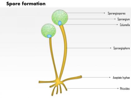
0914 spore formation medical images for powerpoint




Try Before you Buy Download Free Sample Product
 Impress Your
 Audience
 100%
100% Editable
 Save Hours
 of Time


Light The fire with your views. Enhance the combustion with our 0914 Spore Formation Medical Images For PowerPoint.
People who downloaded this PowerPoint presentation also viewed the following :
0914 spore formation medical images for powerpoint with all 10 slides:
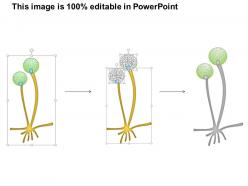
Our 0914 Spore Formation Medical Images For PowerPoint allow you functional freedom. Chop and change as you feel appropriate.




FAQs for 0914 spore formation medical
Fungal spore formation includes endospores, conidia, ascospores, basidiospores, and chlamydospores, each displaying distinct morphological characteristics visible through microscopic imaging techniques. Medical imaging differentiates these through size variations, structural patterns, and cellular arrangements, with pathology laboratories increasingly utilizing advanced microscopy and digital imaging systems to enhance diagnostic accuracy, streamline identification processes, and ultimately deliver faster patient outcomes in clinical mycology.
Medical imaging techniques enhance spore development understanding by providing real-time visualization of morphological changes, enabling precise tracking of cellular differentiation stages, and revealing structural transformations during fungal reproduction cycles. Advanced microscopy methods like confocal and electron imaging allow researchers to observe spore formation mechanisms in pathogenic species, ultimately delivering deeper insights into fungal biology and enabling more targeted antifungal therapeutic strategies.
Spores serve as dormant survival structures for opportunistic pathogens like Clostridium and Bacillus species, enabling persistence in harsh environments and resistance to standard treatments. Through advanced microscopy and imaging techniques, healthcare facilities can identify these oval, refractile structures within bacterial cells, ultimately enhancing diagnostic accuracy and treatment protocols for resistant infections.
Imaging technology assists early diagnosis by detecting characteristic tissue changes, inflammation patterns, and organ involvement before symptoms become severe through CT scans, MRI, and X-rays. These technologies enable physicians to identify infections like anthrax, botulism, and fungal diseases in lungs, brain, and other organs, ultimately delivering faster treatment decisions and improved patient outcomes.
**INPUT**: What specific imaging modalities are most effective for visualizing spores in clinical specimens? **OUTPUT**: Effective imaging modalities for visualizing spores include phase-contrast microscopy, differential interference contrast microscopy, fluorescence microscopy with specific stains, scanning electron microscopy, and advanced confocal techniques. These technologies enhance diagnostic accuracy by enabling rapid identification, improving morphological assessment, and facilitating automated detection systems, with many clinical laboratories finding that combining multiple modalities streamlines pathogen identification while delivering faster diagnostic results.
Medical images aid in tracking spore-based infections by enabling real-time monitoring of contamination patterns, identifying infection clusters, and mapping transmission pathways across healthcare facilities. Through advanced imaging technologies, hospitals and clinics can detect spore concentrations in ventilation systems, patient rooms, and surgical suites, ultimately delivering enhanced infection control protocols and reducing healthcare-associated infection rates.
Medical professionals face challenges including distinguishing spores from similar cellular structures, accounting for staining variability, managing low-contrast imaging, and differentiating between active and dormant spores. These obstacles can be addressed through advanced imaging technologies, standardized protocols, and AI-assisted diagnostic tools, with many laboratories finding that automated analysis systems enhance accuracy while reducing interpretation time, ultimately delivering more reliable diagnoses and improved patient outcomes.
Advanced imaging technologies like high-resolution microscopy, fluorescence staining, digital image analysis, and automated detection systems have revolutionized spore identification by delivering enhanced clarity, precision, and consistency. These innovations enable microbiologists to distinguish subtle morphological differences, reduce human error through automated classification, and accelerate diagnostic processes, ultimately improving patient outcomes in clinical laboratories while streamlining research workflows.
Spore formation significantly complicates antifungal treatment strategies by creating drug-resistant dormant structures that evade standard therapies, requiring extended treatment protocols and combination approaches. Medical imaging reveals spore distribution patterns in organs like lungs and brain tissue, enabling clinicians to target resistant clusters more effectively, ultimately delivering personalized treatment plans and improved patient outcomes in complex mycoses cases.
3D imaging techniques like confocal microscopy, micro-CT scanning, and electron tomography reveal spore surface textures, internal structures, wall thickness variations, and dimensional relationships that traditional 2D methods cannot capture. These advanced visualization approaches enable researchers and medical professionals to analyze spore morphology with unprecedented detail, enhancing pathogen identification accuracy, antibiotic resistance assessment, and diagnostic precision in clinical laboratory settings.
Environmental factors like temperature fluctuations, humidity levels, nutrient availability, and pH variations significantly influence spore formation patterns, timing, and morphology in pathogenic organisms. Advanced imaging techniques, including high-resolution microscopy, fluorescence imaging, and time-lapse photography, can capture these environmental variations by documenting spore development stages, structural changes, and formation density across different conditions, ultimately enabling healthcare professionals to better predict pathogen behavior and enhance diagnostic accuracy.
High-resolution spore imaging enables mycologists and microbiologists to identify species accurately, analyze morphological characteristics, study germination patterns, and document structural variations critical for taxonomic classification. These detailed images enhance research by revealing cellular structures, facilitating comparative studies across fungal species, and supporting diagnostic applications in clinical microbiology, ultimately advancing our understanding of fungal biology and pathogenic mechanisms.
Machine learning algorithms enhance spore morphology interpretation by automating pattern recognition, improving diagnostic accuracy, and standardizing analysis across different imaging systems. These technologies streamline pathogen identification through advanced image processing, feature extraction, and classification models, with many clinical laboratories finding that automated spore analysis delivers faster diagnostic results, reduces human error, and ultimately enables more precise treatment decisions in infectious disease management.
Current imaging methods face limitations including poor resolution for detecting small spores, difficulty distinguishing viable from non-viable spores, challenges with real-time monitoring, and inadequate contrast in dense tissue environments. Advanced solutions like high-resolution MRI, fluorescent tagging technologies, and AI-enhanced imaging algorithms are revolutionizing spore detection by enabling faster identification, improving diagnostic accuracy, and ultimately delivering more precise treatment protocols for infectious disease management.
Public health initiatives benefit from improved spore imaging through enhanced early warning systems, more accurate exposure assessments, and targeted intervention strategies for communities at risk. These advanced techniques enable health departments to monitor allergen concentrations, predict outbreak patterns, and implement preventive measures more effectively, ultimately reducing respiratory illness rates and healthcare costs while protecting vulnerable populations in schools, hospitals, and urban environments.
-
Innovative and Colorful designs.
-
Good research work and creative work done on every template.