


0714 spleen medical images for powerpoint





Try Before you Buy Download Free Sample Product
 Impress Your
 Audience
 100%
100% Editable
 Save Hours
 of Time


Our 0714 spleen Medical Images For PowerPoint are a pure delight. Your audience will soon have that enchanted look.
People who downloaded this PowerPoint presentation also viewed the following :
Content of this Powerpoint Presentation
Description:
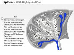
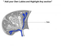
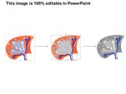
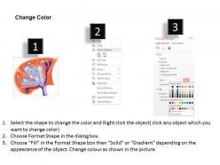
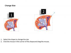
The image illustrates a cross-sectional view of the spleen with its internal structure labeled. The spleen is a part of the lymphatic system and plays a role in both blood filtration and immune system function. The key areas labeled include the capsule, trabecula, vascular sinusoid, red pulp, vein, artery, white pulp composed of primary follicle, marginal zone, periarteriolar lymphoid sheath (PALS), and germinal center.
1. The capsule is the outermost layer that encases the spleen, providing protection and structural support.
2. Trabeculae are extensions of the capsule that branch into the organ, creating a framework for blood vessels and cells.
3. Vascular sinusoids are the channels through which blood flows and are critical for filtering of blood cells.
4. The red pulp primarily functions in filtering and destroying old red blood cells and acts as a reservoir for blood.
5. The vein and artery are the vessels responsible for the inflow and outflow of blood to and from the spleen.
6. The white pulp comprises lymphoid tissue involved in producing and maturing lymphocytes, which are a type of white blood cell. It includes structures like the primary follicle before antigen exposure, the marginal zone that filters antigens from the bloodstream, the periarteriolar lymphoid sheath (PALS) surrounding the splenic arterioles.
7. Finally, the germinal center where B-cell proliferation occurs.
Use Cases:
This detailed image could be utilized across a number of industries for education, training, or information-sharing purposes.
1. Education:
Use: Teaching students about the anatomy and function of the spleen
Presenter: Biology Professor
Audience: Medical and healthcare students
2. Healthcare:
Use: Training healthcare professionals on spleen-related health issues
Presenter: Medical Trainer
Audience: Doctors, nurses, and other healthcare workers
3. Pharmaceutical:
Use: Presenting research and development findings related to drugs affecting the spleen
Presenter: R&D Scientist
Audience: Pharmacologists and healthcare providers
4. Biotechnology:
Use: Discussing advancements in biotechnological applications involving the spleen
Presenter: Biotech Researcher
Audience: Biotechnology professionals
5. Medical Publishing:
Use: Illustrating textbooks or informational booklets on human anatomy
Presenter: Medical Author or Editor
Audience: Readers and learners in the medical field
6. Medical Devices:
Use: Demonstrating how certain medical devices interact with or support spleen function
Presenter: Product Developer
Audience: Investors, healthcare professionals, and regulatory bodies
7. Health Insurance:
Use: Educating about diseases and conditions of the spleen for policy development
Presenter: Health Insurance Educator
Audience: Insurance agents and policy makers
0714 spleen medical images for powerpoint with all 9 slides:
Drive every activity with our 0714 spleen Medical Images For PowerPoint. Get the controls firmly in your hands.




FAQs for 0714 spleen medical
For most spleen stuff, CT with contrast is honestly your best bet - picks up lesions, trauma, sizing issues really well. Ultrasound's great for quick screening since there's no radiation, but bigger patients can be tricky to image. MRI has amazing soft tissue detail, though I feel like it's usually overkill unless you really need that extra info. Trauma cases? CT wins every time for speed and catching bleeds. I'd start with ultrasound for routine checks, then move to CT when you need solid answers. Oh, and ultrasound's perfect for follow-ups too.
So when I'm looking at spleen lesions, benign stuff like cysts usually has clean borders and stays pretty uniform - no enhancement either. Malignant ones? Total opposite. Irregular edges, patchy enhancement, restricted diffusion on MRI. Lymphoma's honestly the worst because it can look totally normal sometimes, which throws everyone off. I always check for enlarged lymph nodes too - malignant lesions love bringing friends. Clinical history matters a ton here. If imaging's unclear, just bite the bullet and get a biopsy. Way better than guessing.
You really can't confirm splenomegaly without imaging - physical exam alone just isn't reliable enough. CT scans and ultrasounds give you actual measurements to compare against normal ranges, which beats palpation every time. Though honestly, if you can feel it during exam, that's already a red flag. The imaging also tells you what's causing the enlargement - could be portal hypertension, lymphoma, infection, whatever. Plus you'll catch complications like infarcts or rupture risk. I always tell people: if you're suspicious clinically, just get the scan. Don't mess around trying to guess.
Honestly, spleen variations mess with your head way more than they should. Accessory spleens are everywhere - I've seen them mimic pancreatic masses so many times. Don't even get me started on wandering spleens that just decide to hang out somewhere random. Size changes constantly with breathing and positioning too, which screws up your measurements. Oh, and early contrast phases? The enhancement looks sketchy as hell but it's completely normal - just heterogeneous timing. Always pull priors if you've got them. Congenital stuff like polysplenia obviously changes everything, but that's pretty rare.
Honestly, ultrasound kinda sucks for spleen stuff. Those ribs get in the way and block your view - so annoying. You'll totally miss smaller lesions that would pop right up on CT or MRI. The bigger issue though? Ultrasound can't really tell you what type of tissue you're looking at. CT shows you those nice contrast patterns, MRI gives you way better characterization. Plus it's super operator-dependent, so results vary wildly between techs. I mean, it's fine for basic screening or checking on something you already know about, but when you actually need to figure out what's going on, just go straight to CT or MRI.
So basically, contrast agents create these density differences that make lesions pop out way more clearly against normal spleen tissue. Without it, you're pretty much guessing on most splenic issues. Different lesions light up at different rates and intensities - cysts vs hemangiomas vs mets all have their own signature patterns. It's actually kind of cool how distinct they are. The contrast shows vascular patterns you'd never see otherwise. Oh, and definitely push for both arterial and portal venous phases if you can - that combo gives you the best shot at nailing down what you're dealing with.
Look, those vascular patterns are basically your cheat sheet for reading spleen images properly. You can trace the splenic artery and vein branches to catch infarcts or masses that you'd totally miss otherwise. Honestly, it's one of those "lightbulb moment" things - seems super obvious after someone shows you, but game-changing for accuracy. The vessel layout also tells you about perfusion issues and systemic blood flow problems. I swear, just spending that extra minute following the vessels around will boost your confidence so much. Worth the time investment for sure.
Look for contrast extravasation first - that's active bleeding and usually means they'll need surgery or IR. Large hematomas are bad news too, especially subcapsular ones since the capsule can rupture later (double rupture thing). Don't stress too much about the grading systems honestly, they're not always reliable for predicting what'll actually happen. Pseudoaneurysms and AV fistulas can pop up days later which is annoying, so you'll need follow-up imaging. Just make sure you document everything clearly and flag the high-risk stuff to trauma right away.
Yeah so age totally matters - older folks usually have smaller spleens with weird echogenicity, plus random calcifications that don't mean anything. But honestly, the underlying conditions are where things get interesting. Portal hypertension makes the spleen huge, blood disorders create this patchy look with random lesions scattered around. Autoimmune stuff? Diffuse enlargement every time. I always check the patient's history and labs first though - makes those confusing images actually make sense. Short sentences help too.
So a few things have really changed the game for spleen imaging lately. High-res CT with multiplanar reconstruction lets you see way more detail in the parenchyma - catches stuff we'd totally miss before. For tricky masses, MRI with diffusion-weighted imaging is honestly incredible for figuring out what you're dealing with. Contrast-enhanced ultrasound works great too, especially for trauma cases where you need real-time perfusion info. The coolest advancement though? AI volumetric analysis that automatically measures everything and tracks changes over time. Pick whatever fits your specific case, but these will definitely boost your confidence when you're stuck on something weird.
Kids' spleens are proportionally huge compared to adults - what looks crazy enlarged might actually be totally normal. Honestly, this gets everyone when they're starting out. Also, you'll see different contrast enhancement patterns and more prominent tissue in pediatric cases. The measurements are obviously way different too, so don't use adult reference ranges. Here's the thing though - kids get splenomegaly from infections way more than adults, who usually have blood cancers or portal hypertension issues. Always think infections first with pediatric cases and use kid-specific standards when you're reading the images.
Motion from breathing is your biggest headache here - breath-hold sequences or respiratory gating help a ton. You'll also get susceptibility artifacts around the splenic flexure from bowel gas (adjust shimming or pick sequences that aren't as picky about field inhomogeneities). Flow artifacts from vessels nearby? Flow compensation gradients or saturation bands usually do the trick. Honestly, getting patient positioning right upfront saves you way more time than trying to salvage crappy images later. Oh, and don't forget vascular flow artifacts - they're annoying but pretty fixable with the right sequence tweaks.
CT with contrast is definitely your go-to here - skip the plain films, they won't tell you much. You'll want to measure spleen size and check for accessory spleens that might be hiding. The relationship to the pancreatic tail matters too since that's where things get tricky. Look for any masses or weird vascular anatomy that could bite you during surgery. Honestly, the imaging is only half the story though. Always pair it with platelet counts because that combo gives you the real picture of what's going on functionally. These findings help you plan your approach and prep patients for potential complications.
Fat tissue really messes with ultrasound waves - it's like trying to see through a thick blanket. Your image quality takes a huge hit because the waves can't penetrate well and you get tons of scatter. Plus the spleen's sitting way deeper, so those echoes coming back are super weak. I'd switch to a lower frequency probe for better penetration, but yeah, you'll lose some sharpness. Harmonic imaging helps when you've got it available. Honestly though? If the ultrasound looks terrible, just go straight to CT or MRI instead of wasting time fighting with crappy images.
Dude, AI for spleen imaging is actually pretty incredible. It catches those tiny abnormalities you'd totally miss on a busy day. The algorithms spot early lesions and measure splenic volume way more precisely than we can eyeball it. Plus they pick up texture changes that scream lymphoma or infarction - honestly sometimes better than I do after my third cup of coffee. Speed's the best part though. You get consistent measurements in seconds instead of sitting there with calipers forever. It also cuts down on that annoying variability between different radiologists reading the same scan. Definitely try it on your trickier hematologic cases.
-
Top Quality presentations that are easily editable.
-
Professional and unique presentations.