


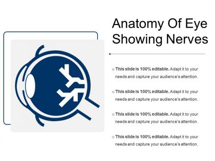
Anatomy of eye showing nerves




Try Before you Buy Download Free Sample Product
 Impress Your
 Audience
 100%
100% Editable
 Save Hours
 of Time


Get folks interested in achieving common goals with our Anatomy Of Eye Showing Nerves. Be able to involve everyone.
People who downloaded this PowerPoint presentation also viewed the following :
Anatomy of eye showing nerves with all 5 slides:
Groom them for the future with our Anatomy Of Eye Showing Nerves. They will develop into a capable bunch.




FAQs for Anatomy of
The optic nerve's primary functions include transmitting visual information from retina to brain, processing light signals into neural impulses, enabling depth perception, coordinating binocular vision, and facilitating peripheral vision detection. These neural pathways enhance visual processing by integrating sensory data, coordinating eye movements, and delivering comprehensive sight capabilities, with healthcare professionals and educational institutions finding that understanding these mechanisms ultimately streamlines diagnostic approaches and treatment outcomes.
The retina converts light into electrical signals through photoreceptors, while the optic nerve transmits these signals to the brain for processing and interpretation. This partnership enables visual perception by transforming optical information into neural data that the visual cortex processes, ultimately delivering the complex visual experiences essential for navigation, recognition, and decision-making in daily activities.
Ganglion cells serve as the eye's primary output neurons, collecting processed visual information from bipolar and amacrine cells and transmitting it to the brain through their axons, which form the optic nerve. These specialized cells enable critical visual functions like motion detection, contrast sensitivity, and spatial processing, with different ganglion cell types facilitating distinct aspects of vision, ultimately delivering comprehensive visual perception and enabling rapid response to environmental changes.
The optic nerve differs from other cranial nerves through its unique myelination by oligodendrocytes rather than Schwann cells, larger diameter fibers, and direct brain tissue extension rather than peripheral nerve structure. This distinctive anatomy enables enhanced visual signal transmission and processing capabilities, with the optic nerve's specialized architecture ultimately delivering superior bandwidth for complex visual information compared to traditional peripheral cranial nerves.
Common optic nerve pathologies include optic neuritis, glaucoma, optic nerve atrophy, papilledema, and ischemic optic neuropathy. These conditions impact vision by causing visual field defects, decreased visual acuity, and color vision abnormalities, with many healthcare institutions finding that early detection through comprehensive ophthalmologic screening enables better treatment outcomes and preserves patients' functional vision.
The visual pathway facilitates image processing through the optic nerve, optic chiasm, lateral geniculate nucleus, and visual cortex, creating a systematic information relay system. This neural architecture enables rapid signal transmission and parallel processing, with healthcare institutions and medical training programs finding that understanding these pathways enhances diagnostic accuracy and treatment outcomes.
Eye nerves adapt to light changes through pupillary reflexes, photoreceptor sensitivity adjustments, and neural pathway modulation. The optic nerve coordinates with sympathetic and parasympathetic systems to regulate pupil size, while retinal cells shift between rod and cone dominance, ultimately enabling seamless vision transitions from bright office environments to dimly lit spaces.
The parasympathetic system constricts pupils through the oculomotor nerve and enhances near vision, while the sympathetic system dilates pupils via cervical ganglia and prepares for distance focus. These complementary systems enable dynamic visual adaptation by regulating pupil size, lens accommodation, and tear production, ultimately delivering optimal vision across varying light conditions and distances.
Advanced imaging techniques for studying eye nerves include optical coherence tomography (OCT), adaptive optics scanning laser ophthalmoscopy, confocal scanning laser microscopy, and high-resolution magnetic resonance imaging. These technologies enhance diagnostic precision by delivering detailed structural analysis, real-time visualization capabilities, and comprehensive nerve fiber mapping, with ophthalmology practices finding that improved imaging ultimately streamlines patient care and enables earlier intervention strategies.
Neurotransmitters facilitate eye neural communication by enabling signal transmission between photoreceptors, bipolar cells, horizontal cells, amukrine cells, and ganglion cells through chemical synapses. These specialized molecules, including glutamate, GABA, and dopamine, streamline visual processing by modulating light-dark adaptation, contrast enhancement, and motion detection, ultimately delivering precise visual information to the brain's processing centers.
The pupillary light reflex serves as a critical diagnostic tool for evaluating optic nerve function, as it directly tests the neural pathway from retina to brainstem via the optic nerve. An abnormal or absent pupillary response often indicates optic nerve damage, retinal disorders, or brainstem lesions, enabling clinicians to quickly assess neurological health and differentiate between various conditions affecting visual processing and nerve transmission pathways.
Glaucoma affects eye nerve function by increasing intraocular pressure, which damages the optic nerve fibers and disrupts signal transmission to the brain. This progressive condition initially impairs peripheral vision through nerve cell death, ultimately leading to tunnel vision or complete blindness if untreated, with early detection and pressure management being critical for preserving visual function.
Environmental factors impact eye nerve health through exposure to UV radiation, blue light from digital devices, air pollution, and chemical irritants that can cause oxidative stress and inflammation. These factors can lead to conditions like glaucoma and optic neuropathy, with many ophthalmologists finding that protective measures like UV-filtering eyewear and regular eye exams help maintain optimal nerve function and prevent long-term damage.
The eye's nervous system enables depth perception through binocular vision processing, retinal disparity analysis, convergence mechanisms, and motion parallax detection. These neural pathways work by comparing images from both eyes, calculating distance variations, and integrating movement cues, ultimately delivering enhanced spatial awareness that enables precise navigation, improved hand-eye coordination, and better environmental assessment across various professional and personal applications.
Optic nerve injury significantly impacts visual function through reduced acuity, visual field defects, color perception changes, and potential complete vision loss in severe cases. While recovery varies depending on injury extent and timing of intervention, many patients find that early rehabilitation programs, adaptive technologies, and comprehensive ophthalmologic care can optimize remaining vision and enhance quality of life outcomes.
-
Great experience, I would definitely use your services further.
-
Excellent template with unique design.