


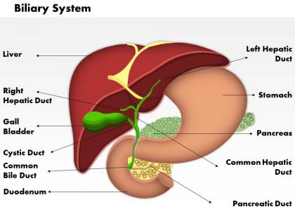
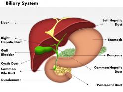
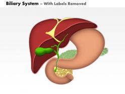
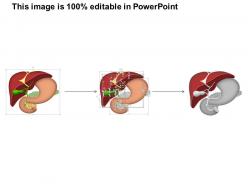
0514 biliary system medical images for powerpoint




Try Before you Buy Download Free Sample Product
 Impress Your
 Audience
 100%
100% Editable
 Save Hours
 of Time


Take a different approach with our 0514 Biliary System Medical Images for PowerPoint. Initiate anew course with your thoughts.
People who downloaded this PowerPoint presentation also viewed the following :
0514 biliary system medical images for powerpoint with all 11 slides:
Funnel your thoughts accurately with our 0514 Biliary System Medical Images for PowerPoint. They will flow along the correct channel.
FAQs for 0514 biliary system medical
For most biliary stuff, ultrasound is your go-to for screening - catches gallstones really well. MRCP comes next when you need detailed ductal views, especially for suspected obstruction. It's non-invasive which patients love. ERCP is the big gun - more invasive but you can actually do something therapeutic if you find stones or strictures. CT with contrast works too for masses and ductal dilation. Honestly, ultrasound first then MRCP covers like 90% of cases. Just remember bowel gas will mess with your ultrasound at the most inconvenient times possible.
So MRCP is basically MRI but dialed in specifically for looking at bile ducts. Regular MRI shows your liver fine, but MRCP cranks up this T2-weighting thing that makes bile glow bright white while everything else stays dark. Makes bile ducts pop like crazy - you can see stones, blockages, all that stuff without needing contrast or any invasive procedures. Honestly it's pretty wild how clear the images are. If your doc's checking for gallbladder or bile duct problems, definitely push for MRCP over regular liver MRI. Way better for catching ductal issues.
Ultrasound's your go-to for anything biliary. Works great for spotting gallstones and checking if bile ducts are dilated or the gallbladder wall looks thick. You can literally do Murphy's sign while you're scanning, which is honestly pretty satisfying when it's positive. Plus you get real-time imaging right at bedside - no radiation, doesn't cost much, and it's quick. Downside? Fat patients and bowel gas will mess with your views sometimes. But yeah, always start with US for biliary stuff, then if you need better duct detail you'll have to move up to MRCP or ERCP.
So basically you order it when someone's got RUQ pain, jaundice, or funky liver enzymes - think gallbladder problems or bile duct issues. Murphy's sign? Definitely time for imaging. Classic stuff like cholecystitis, stones in the bile duct, or ruling out biliary pancreatitis. Post-op complications are another big one - bile leaks can be sneaky. Oh, and chronic things like PSC obviously need monitoring. Honestly, ultrasound is your friend for gallbladder stuff first, then you can get fancy with MRCP or ERCP if needed. Pretty straightforward once you get the hang of it.
Ultrasound is still the best first choice - you'll see those bright spots with shadowing behind them. CT scans can totally miss cholesterol stones since they look just like bile, but calcified ones show up bright white. MRI's pretty cool because stones look like dark holes on T2 images, plus you get amazing views of all the bile ducts. ERCP will show filling defects if someone's going that invasive route. Honestly ultrasound wins for screening - it's cheap and fast. Oh, and tiny stones or that sludgy stuff doesn't always cast shadows consistently, which can be annoying.
So you're basically looking for dilated bile ducts above the blockage - they'll show up as these branching tubes that are way bigger than they should be. Common bile duct over 6mm is suspicious (10mm if they've had their gallbladder out). Sometimes you can actually see what's causing it - stone, stricture, whatever. The gallbladder usually gets swollen too, especially with downstream blockages. That's the Courvoisier thing when it's not painful. Oh and don't forget to check labs - biochemical obstruction doesn't always match what you see on imaging, which honestly trips people up more than it should.
So for cholangitis on CT, ductal dilatation is your biggest clue - usually jumps out pretty quickly. Look for wall thickening around those ducts too. Enhancement patterns can be super helpful, especially that periductal stuff around the porta hepatis. Sometimes you'll catch fluid collections or even abscesses if it's gotten nasty. The hepatic parenchyma might look weird too. Honestly, I used to miss subtle cases until I got into a routine of always checking ductal caliber systematically - now it's way easier to spot. Just don't expect every case to be textbook obvious, some are pretty sneaky at first glance.
MRCP is gonna be your go-to here - shows that classic "abrupt cutoff" where the duct just suddenly narrows. You'll see focal wall thickening and strictures that look irregular, plus upstream biliary dilatation. CT can be tricky though, sometimes just shows subtle wall enhancement that's super easy to miss. The big thing is asymmetric involvement vs. the smooth, concentric narrowing from benign strictures. Also check for lymphadenopathy around the porta hepatis. Honestly, if you're seeing this stuff in an older patient, definitely push for tissue sampling or closer follow-up imaging. Better safe than sorry with these cases.
So these congenital biliary things have pretty telltale signs once you know what to look for. Choledochal cysts show up as dilated intrahepatic ducts - super obvious on MRCP or CT. With biliary atresia, you'll see absent or threadlike extrahepatic ducts plus progressive dilation inside the liver. Caroli disease has that weird "central dot sign" on MRI where the portal vein sits right in the middle of the dilated ducts. MRCP beats CT hands down for ductal detail, honestly. Quick recognition matters since most need surgery, especially in kids where timing can make or break the outcome.
Small ducts are the worst - anything under 2-3mm basically disappears on CT or MRI. MRCP misses tiny stones under 3mm too. Image quality gets terrible when patients can't hold their breath (which happens more than you'd think). Here's the annoying part: benign and malignant strictures look exactly the same on imaging. You're seeing structure but not function, so who knows if things are actually draining well? I always combine different scans and check labs. The clinical picture tells you way more than any single image will.
So with ERCP you're literally injecting contrast right into the bile ducts through the ampulla - gives you insane detail on strictures, stones, all that anatomy stuff. But here's the cool part: you can actually fix things during the same procedure. Sphincterotomy, pull out stones, drop in stents. It's wild how much you can get done in one go. MRCP works fine for screening I guess, but when you need to see everything clearly AND actually treat whatever's wrong? ERCP's your move. Way more invasive obviously, but sometimes you gotta do what works.
Honestly, just think about what you're actually trying to figure out first. Ultrasound's your friend for basic stuff - stones, whether ducts look dilated. Super quick and won't break the budget. CT gives you the whole picture if you need to see everything, but yeah, radiation sucks. MRCP is probably your best bet for really detailed duct imaging without worrying about contrast reactions, though good luck if your patient's claustrophobic or can't lie still for 45 minutes. ERCP's obviously the gold standard when you actually need to do something, not just look. Really comes down to matching what you pick with what you need to know and whether your patient can handle it.
Look at where the dilation stops - that's your answer. Dilated ducts inside the liver but normal ones outside? You've got intrahepatic obstruction. Both dilated? Then it's extrahepatic, probably ampulla or pancreatic head stuff. MRCP will save your life here, honestly way better than trying to guess. You can trace the CBD on ultrasound too if you're patient enough. The spot where dilated meets normal caliber - that's literally pointing to your blockage. Makes sense once you think about it that way.
So there's some pretty cool stuff happening with biliary imaging right now. AI-enhanced MRCP is way better at catching those sneaky ductal issues we used to miss - honestly the image quality blows my mind. EUS with contrast is getting super precise for strictures and masses too. The newer cholangioscopy systems? Total game changer. We're talking direct visualization that would've seemed like sci-fi a few years back. Best part is you get all this detailed anatomical info without putting patients through anything too invasive. Definitely worth chatting with your rad team about what they've got available - some places are ahead of the curve on this stuff.
Look, it really comes down to what you're hunting for. Acute cholangitis? Focus on ductal dilatation and wall thickening on MRCP. Post-surgical cases are different - you're checking anastomotic sites for leaks. Stone disease is all about filling defects and caliber changes, though honestly those tiny stones can be ridiculously sneaky sometimes. Malignancy workups need careful stricture assessment and mass evaluation. Emergency presentations require quick obstruction checks. Surveillance studies let you take your time with detailed anatomy. Just match your approach to the clinical question - sounds obvious but you'd be surprised how often people miss that.
-
Very unique, user-friendly presentation interface.
-
Best Representation of topics, really appreciable.