

0514 cross section of the spinal cord medical images for powerpoint





Try Before you Buy Download Free Sample Product
 Impress Your
Audience
 100%
Editable
 Save Hours
of Time


Believe in the day with our 0514 Cross Section Of The Spinal Cord Medical Images For Powerpoint. Your thoughts will experience a fresh dawn.
People who downloaded this PowerPoint presentation also viewed the following :
Content of this Powerpoint Presentation
For a human body, the spinal cord is one of the crucial organs that coordinates action and reaction based on the signals from the brain and the sensory organs. Because the brain does not directly connect with all parts of the body and it must initiate quick response under life threatening or even life-sustaining processes, the spinal cord acts as the reliable support to ensure there is no delay.
An organ that is cylindrical in shape running from the hindbrain to the tailbone, this other better half of the Central Nervous System becomes a crucial subject of understanding for medical students and the laymen alike. Learning about this vital organ and its functions followed by its protective care needs to be the duty of every health-conscious individual. In the current section, our focus will be on understanding the anatomy of spinal cord, a body part the medical profession reveres. It is a common subject of interest for ophthalmologists, nephrologists, orthopedics, neurologists, and even the general physicians. SlideTeam has designated this presentation template to portray the anatomy of a spinal cord. It is useful for medical students and relevant audiences to have a visual understanding of the spinal cord’s cross-section. This PPT Preset is color-coded and well-labeled that makes it ready to display and use as a reference. Learn more with the PPT Template below:
Template 1: Cross-Section of the Spinal Cord Medical Image PPT Template
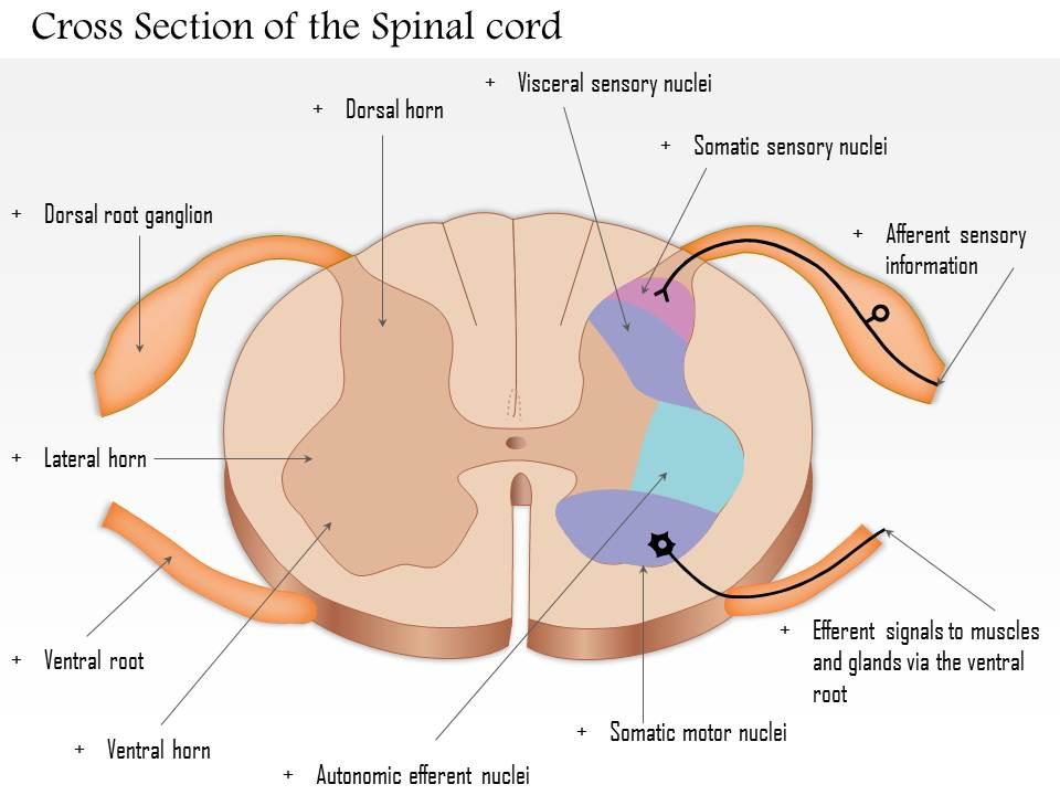
Get this PPT Diagram of the spinal cord’s cross-section to demonstrate its inner anatomy. Proper labeling has also been provided in this colored diagram of the spinal cord. Illustrate the process of receiving sensory information via afferent nerves (sensory nerves with axons in the spinal cord) to signaling respective muscles and glands, via efferent nerves(motor nerves with dendrites in the spinal cord). Sectioning of the inner, mid, and outer parts of the spinal is also clear in this PPT Design and color-coding will help you point out their specific role in their information interchange process during your presentation. Lateral Horn, Dorsal horn, and Ventral horn among other parts of the cross-section diagram are also vivid and will add to your detailed discussion of the spinal cord's internal structure.
Educate your audience on the crucial roles of individual sections of the spinal cord using this PPT Layout. As the presentation template is fully editable, you can add/remove text, re-color, or re-orient elements and text. In this way, you can use this diagram and make it anything between simple and detailed.
PS: Here’s a ready-made diagram of the lateral view of a spinal cord to indicate the detailed structure of the 31 neurons that radiate from it.
0514 cross section of the spinal cord medical images for powerpoint with all 11 slides:
Rigorous design has gone into making sure our 0514 Cross Section Of The Spinal Cord Medical Images For Powerpoint collection is the finest one available today. Thousands of designs ready at a click.


FAQs for 0514 cross section of the spinal cord medical
So you'll mainly see gray matter in the center - it's that butterfly shape everyone talks about, and honestly once you notice it you can't unsee it. White matter surrounds it with all the tracts going up and down your spine. The gray stuff has dorsal and ventral horns with cell bodies. There's also this tiny central canal running through the middle, plus meninges around everything. My advice? Focus on spotting that gray-white contrast first. Makes everything else way easier to identify. Oh and don't forget about the CSF space - shows up pretty clearly usually.
Okay so the gray matter shape is your biggest clue here. Cervical ones have that butterfly look with big anterior horns - they're also the largest overall. Thoracic sections? Way smaller and rounder, plus they've got those lateral horns that honestly make them super easy to ID once you know what you're looking for. Lumbar gets big again with huge anterior horns for all the leg muscles. Sacral's tiny and circular. The gray-to-white ratio shifts a lot between levels too, which helps. I'd start with cervical and lumbar since they're obvious, then tackle thoracic after.
Looking at spinal cord cross-sections can really tell you what's happening neurologically. Demyelination patterns scream MS, while cavities usually mean syringomyelia. Asymmetric lesions? Think tumors or strokes. Location's huge too - cervical problems mess with both arms and legs, but thoracic issues typically leave the arms alone. Some patterns are honestly so textbook they practically diagnose themselves. You can measure cord shrinkage or count lesions to see how bad things are getting. The real trick though is matching where you see the damage with what your patient's actually experiencing. That's where everything clicks.
Looking at spinal cord sections, the gray matter totally changes shape depending where you are. Cervical and lumbar areas have these massive anterior horns - that's where all the motor neurons for arms and legs live. White matter does the reverse, getting thicker as you go up because more tracts are joining in. Cervical sections look like chunky butterflies honestly, while thoracic ones are way more compact and oval. Sacral sections are tiny but gray matter takes up more space proportionally. Pro tip: always check what level you're looking at first - it'll instantly tell you the gray-to-white ratio you should expect.
Dude, those cross-section images are everything for spinal cord injuries. They literally show you exactly where the damage is - which pathways got messed up, what tissue looks funky. It's wild how precise they are. Different spine levels control different stuff, so seeing the exact location tells you what functions might be screwed. Doctors use them to track if someone's getting better too. Oh and definitely match up what the scans show with actual symptoms - sometimes there's weird disconnects there. Makes planning treatment way more targeted instead of just guessing.
MRI's definitely your best bet for spinal cord imaging - the soft tissue contrast is just incredible. CT works great too, especially for bone stuff or when someone can't handle the MRI machine (claustrophobia, pacemakers, whatever). Oh, and CT myelography is a thing where they shoot contrast into the spinal canal. Histological sectioning is still gold standard for research but that's obviously post-mortem territory. T2-weighted sequences are money for pathology - lesions, edema, all the anatomical details pop right out. Honestly can't go wrong starting there.
Oh man, spinal cord cross sections are actually super useful for tracking disease progression! You can literally see how the cellular structure breaks down over time - like demyelination in MS or when motor neurons die off in ALS. The inflammatory patterns are wild when you zoom in. Honestly, it's kind of morbid but fascinating? Researchers use these "before and after" shots to find biomarkers and figure out which cells get hit hardest. If you're doing anything with spinal pathology, you'd be crazy not to include cross-sectional analysis. It's one of those techniques that just makes sense.
So here's the thing - nerve root pathways basically give you a roadmap to figure out where symptoms are actually coming from. Like, that toe numbness? Could totally be from the lower back, not the foot. You can trace numbness, weakness, or pain back to the exact nerve that's acting up by following these pathways on your scans. Super useful for planning surgeries or figuring out injection sites. Always double-check that your imaging matches what the patient's telling you though - sometimes things don't line up perfectly and you'll need to dig deeper.
Dude, the spinal cord cross-section is actually super cool once you get it. Gray matter does all the local processing stuff - think reflexes and immediate responses. White matter? That's your highway system carrying signals up and down. Motor pathways stick to specific spots (corticospinal tracts hang out laterally), sensory paths have their own neighborhoods, and autonomic functions cluster together too. Here's the kicker - you can literally predict what'll go wrong based on where the damage hits. I used to hate anatomy until this clicked for me. Next time you're looking at imaging, try spotting the major tracts first. Makes everything way clearer.
Looking at spinal cord cross-sections is honestly amazing for spotting degenerative diseases. Gray matter loss jumps right out at you, plus white matter damage and cell density changes that match up with ALS, MS, or spinal muscular atrophy. Compare sections from different spine levels and you can literally track how the disease spreads. Inflammation patterns are super helpful too - they help you tell diseases apart. Oh, and if you're dealing with motor neuron stuff, check the ventral horn first. That's where things go downhill fastest. You'll see gliosis and demyelination pretty clearly there.
Oh man, color-coded models are seriously a lifesaver for spinal cord stuff. Each color represents different parts - gray matter, white matter tracts, specific nuclei. No more staring at those awful grayscale images trying to figure out what's what. Students always get it way faster with the colored versions too. You can actually follow pathways between different spinal levels without losing your mind. Plus spotting lesions becomes so much easier when everything's clearly marked. I swear, once you try overlaying a color template on cross-sections, you'll wonder how you ever managed without it. Total game changer.
You'll mostly run into motion artifacts - they look blurry and streaky. Hardware like screws creates susceptibility artifacts with signal dropout and distortion around the metal. CSF flow artifacts are annoying, they ghost in the phase-encoding direction. Watch for truncation artifacts too - those alternating bright/dark lines near high-contrast areas can really throw you off if you're not paying attention. Chemical shift shows up as tissue displacement, usually at fat-water interfaces. Honestly, the patterns and locations make them pretty recognizable once you know what to look for. Just double-check against patient history and hardware placement so you don't mistake artifacts for actual pathology.
Dude, cross-sectional imaging is literally a lifesaver for surgical planning. Before you even touch the patient, you can see exactly where the pathology is and map out all the critical stuff - nerve roots, blood vessels, the whole deal. It helps you figure out the best approach and entry point too. I mean, trying to plan complex spine cases without these images would be like operating blindfolded honestly. Oh and it's clutch for picking the right instrument sizes ahead of time. My attending always says to pull up those cross-sectional views during pre-op because surprises in the OR are never fun.
Yeah, age is way more important than sex when you're looking at spinal cord samples. Older people show some pretty obvious changes - white matter gets less dense, you'll see demyelination, gray matter looks different with fewer motor neurons visible. Sex differences? Honestly they're pretty subtle, maybe just slight size variations but nothing that really stands out. The aging stuff is so much more noticeable and actually matters clinically. Oh, and definitely check the age on specimens first - it's a lifesaver for figuring out if what you're seeing is just normal aging or actual pathology.
Netter's atlas is literally a lifesaver for this stuff - saved me so many times. Also check out University of Michigan's online spinal cord atlas, it's free and super detailed. Kenhub has these interactive modules that are actually pretty decent, though I found AnatomyZone hit or miss sometimes. What really helped me was doing those tracing exercises over and over. Boring but it works. Try looking at the same cross-section in different atlases side by side - you'll start seeing patterns in the gray matter way faster. Start with cervical sections first since they show the most detail.
-
Design layout is very impressive.
-
Colors used are bright and distinctive.