

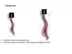
0514 spinal cord lateral view medical images for powerpoint




Try Before you Buy Download Free Sample Product
 Impress Your
 Audience
 100%
100% Editable
 Save Hours
 of Time


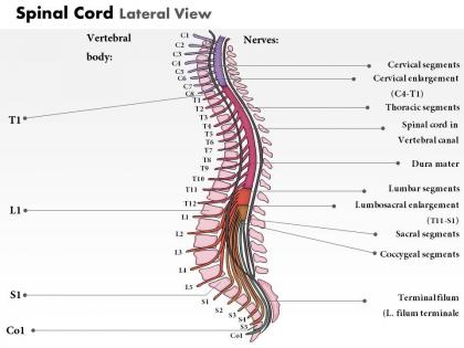
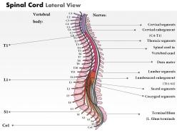
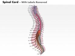
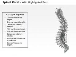
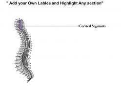
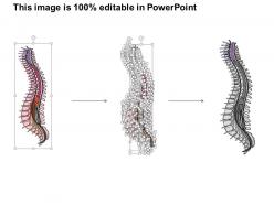
Belonging to a medical background you yourself would understand how important it is to have a well detailed out diagram with all the proper labeling. Therefore our experts have designed for you 0514 spinal cord lateral view medical images for PowerPoint. This PowerPoint presentation visual has a very clear depiction of the backbone i.e. the spinal cord of the human body with all the clear marking of its various parts. With so much and more attached to your medical PowerPoint presentation example, you as a presenter simply need to speak your part and let the remaining be governed by this human spinal cord PowerPoint presentation example. The spinal cord is a long, thin mass of bundled neurons that carries information through the vertebral cavity of the spine beginning at the medulla oblongata of the brain on its superior end and continuing inferior to the lumbar region of the spine. Our 0514 Spinal Cord Lateral View Medical Images For PowerPoint come with no caveats. They have no covert agenda.
People who downloaded this PowerPoint presentation also viewed the following :
0514 spinal cord lateral view medical images for powerpoint with all 9 slides:
Get more than a penny for your thoughts. Increase earnings with our 0514 Spinal Cord Lateral View Medical Images For PowerPoint.

FAQs for 0514 spinal cord lateral view medical
So basically you want this when someone's got neurological stuff going on - weakness, numbness, that shooting pain down their leg. It's perfect for seeing disc herniations, spinal stenosis, or if the cord's getting squeezed. Also great for tumors, infections, or planning surgery. The sagittal view is honestly so much better than axial cuts for seeing how everything lines up across multiple levels - you can actually see what's happening to the discs and nerves. Oh, and definitely get it for trauma cases. If your patient's got unexplained neuro symptoms that might be spine-related, this should be near the top of your list.
So lateral views show you the whole spine from the side - great for spotting alignment issues and disc problems along the entire length. Axial views are those cross-sectional cuts that let you see exactly what's going on inside the cord at each level. Honestly, I always think of it like looking at a house from outside versus checking out the floor plan of each room. You really need both though. I'd start with lateral to get the overall picture, then jump to axial when you find something sketchy that needs a closer look.
Lateral spinal MRIs are perfect for catching disc herniations and spinal stenosis - you get amazing views of those disc spaces. They're also great for vertebral fractures, tumors, infections, and degenerative stuff like spondylolisthesis. Honestly, if I could only pick one view for spine issues, it'd probably be lateral. You can spot cord compression easily, plus any weird curves in the spine. The sagittal shots give you this nice big-picture view across multiple levels that you just don't get with axial cuts. I always start there before getting into the detailed axial analysis - it's like getting the lay of the land first.
Motion from the patient moving or breathing blurs everything, which sucks when you need sharp detail for spinal cord issues. CSF flow creates these ghost images that can totally look like real lesions - honestly one of the most frustrating things to deal with. Metal implants cause signal dropout and distortion. You'll also get chemical shift that moves tissue boundaries around, making measurements pretty unreliable. The main thing is learning to spot these artifact patterns so you don't confuse them with actual pathology. Always check against clinical symptoms, and don't hesitate to repeat with different sequences if the artifacts are screwing up your read.
T2-weighted sequences are gonna be your go-to for lateral spinal imaging - they show cord detail and pathology really well. Sagittal STIR is clutch too, picks up edema that T2 sometimes misses. Stick with 3-4mm slice thickness and minimal gaps. Patient positioning is huge here - honestly, even tiny movements will completely wreck your images, which is so frustrating. T1 post-contrast helps if you're looking for enhancement, but most of the time T2 and STIR do the heavy lifting. Just make sure you're covering your whole area of interest. The contrast on T2 is usually pretty solid for most pathology.
Dude, positioning is everything for lateral spine MRIs. If the patient's not perfectly straight and parallel to the table, you'll get oblique cuts instead of true sagittal views. That rotation screws up your anatomy visualization big time. I've literally seen disc herniations look way worse because someone rushed the setup. The localizer images are your best friend here - spend that extra minute checking before you run the full sequence. Nothing's more annoying than having to repeat an entire study because you didn't nail the positioning from the start. Poor alignment can make normal stuff look pathological too.
Honestly, MRI is your best bet for anything involving the actual spinal cord. The soft tissue detail is just so much better - you'll catch stuff like demyelinating lesions, cord swelling, tumors that CT totally misses. CT's great if you're worried about fractures or need something fast in trauma, but for inflammatory conditions or degenerative changes? MRI wins every time. You can actually see CSF flow and subtle compression. I mean, if your patient can handle lying still for the longer scan and it's not an emergency situation, definitely go MRI for any suspected cord problems.
Honestly, the imaging has gotten so much better lately. 3T scanners give you way sharper detail now. DTI is the real winner though - it shows white matter tracts and catches subtle injuries that regular sequences totally miss. Motion artifacts used to be such a pain for sagittal views, but the newer fast spin-echo sequences handle that way better. Better coils help with signal-to-noise too. If you're not using DTI yet, you really should push for it. The diagnostic pickup rate for cord stuff goes up big time. I think some places are still stuck on old protocols which is kind of frustrating.
Hey! So for lateral spine MRIs, I always check three main things. Cord diameter first - it should stay pretty consistent except where it naturally tapers at the conus. Then look at the contour for any weird angulation or kyphotic changes that might mean compression. Signal intensity is huge too, especially T2 changes that show edema or myelomalacia you'd miss on sagittal cuts. Honestly, lateral views don't get enough love but they're clutch for seeing anteroposterior relationships. Oh, and if there's any hint of instability from the history, definitely push for flexion-extension views. Makes such a difference in catching dynamic stuff.
Honestly, contrast makes a huge difference for catching inflammatory stuff and tumors on spinal MRI. Gadolinium lights up areas where blood vessels are acting weird or tissue barriers break down. You'll miss active MS plaques and some spinal cord tumors without it - they can look identical to normal tissue on regular T1. I've seen so many cases where the lesion was basically invisible until contrast kicked in. Kind of frustrating when you're squinting at scans thinking "something's off here" but can't pin it down. If your patient can handle the contrast and you're worried about inflammation or cancer, definitely worth pushing for it.
Lateral views show you how much cord is involved vertically - you can see lesions running along the length and get their cranial-caudal extent. Axial cuts give you the cross-sectional story: is it hitting dorsal columns, lateral funiculi, gray matter? I always check both since they tell different parts of the story. Measuring lesion length on lateral is super helpful for distinguishing tumefactive demyelination from actual tumor. Cross-sectional views answer what's affected at each level. Honestly, don't make your call until you've looked at both planes - they're like puzzle pieces that fit together.
You're missing way too much with just sagittal - that's the biggest issue. Cross-sectional axial cuts show you the real story for cord compression, nerve root problems, all that stuff. Sagittal's fine for alignment and basic cord shape, but anatomical overlap hides smaller findings. Coronal helps too. I mean, I used to rely more on sagittal views but honestly don't anymore. You'll catch so much more pathology with at least two planes. If the clinical picture's complicated, go for all three. Trust me on this one.
Dude, those lateral MRIs are a game-changer for spine surgery prep. The sagittal view shows you everything - spinal alignment, disc spaces, where the cord's getting squeezed. You'll catch ligamentum flavum thickening and central stenosis that axial cuts sometimes miss. Plus they help figure out your surgical angles and whether you need hardware. I mean, who wants to get surprised in the OR, right? Always look at these with your axials before scheduling anything. Makes planning laminectomies or fusions so much cleaner when you can see the whole picture first.
Look, lateral views alone won't cut it for most spinal cord stuff. You'll want sagittal T2 and T1 sequences - those are your go-to for spinal pathology. Axial cuts through the problem area are clutch, especially for cord compression or lesions. Trauma cases? Might need flexion-extension views. Honestly, I always correlate with clinical findings first. If you're thinking infection or tumor, definitely add contrast. Don't be shy about ordering a full spine protocol if something seems weird - better safe than sorry.
Lateral view is clutch for seeing compression and how messed up the alignment actually is. Axial cuts show you what's damaged but miss the big picture - you need sagittal to see if vertebrae are sliding around or if there's canal narrowing. Honestly it's way more useful for trauma than most people realize. You can spot ligament tears, see how much room the cord has left, and figure out if multiple levels got hit. The lateral basically tells you how unstable everything is, which is what drives your OR decisions. Obviously still check your neuro exam, but that view guides whether someone needs emergency surgery or not.
-
Topic best represented with attractive design.
-
Content of slide is easy to understand and edit.