


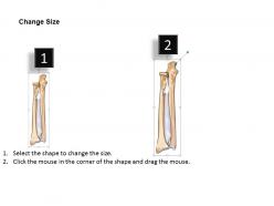
0514 radius anterior view medical images for powerpoint




Try Before you Buy Download Free Sample Product
 Impress Your
 Audience
 100%
100% Editable
 Save Hours
 of Time


Don't let half truths cause any damage. Our 0514 Radius Anterior View Medical Images For PowerPoint give access to the full gen.
People who downloaded this PowerPoint presentation also viewed the following :
0514 radius anterior view medical images for powerpoint with all 10 slides:
Our 0514 Radius Anterior View Medical Images For PowerPoint are exclusively crafted. They can be alluringly awesome.





FAQs for 0514 radius anterior view medical
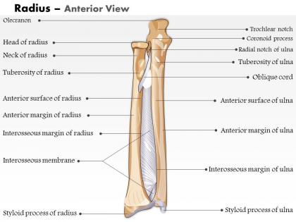
On that anterior radius view, look for the radial head and neck up top, plus the tuberosity. The shaft runs down to the styloid process at the bottom. You'll spot where it connects with the ulna too - honestly those connection points are where most fractures happen anyway. Check the bicipital tuberosity on the medial side, it shows up pretty well usually. Watch for any breaks in the bone outline or weird spacing in the joints. Radial head injuries are super common so definitely focus there first when you're going through the images.
So those anterior view radius images are super helpful for catching fractures you'd totally miss on just lateral views. Hairline cracks, displacement, weird angulation - they all show up way clearer from this angle. I swear half the cases I've seen, the AP view was what actually caught it. You can measure shortening better too and see if bone fragments are overlapping. Oh, and don't forget to check those radiocarpal joints while you're at it. Honestly though, always look at both views together - never trust just one when you're hunting for radius fractures.
The 0514 radius anterior view is solid for catching distal radius fractures - Colles' and Smith's fractures show up really clearly. You'll also spot radial head fractures and any joint space narrowing from arthritis. Bone alignment is super obvious from this angle, plus you can catch those sneaky cortical breaks that are easy to miss. Actually, I've always found fractures just jump out at you on this view. Oh, and definitely pair it with the lateral - you need both angles or you're gonna miss stuff. The anterior positioning just gives you that clean perspective you can't get otherwise.
Hey! So for those anterior radius views, put the radial head up top and make sure that styloid process goes toward bottom right. Thumb side goes right, ulnar side left - honestly sounds super basic but I swear people mess this up constantly lol. You want that radial tuberosity visible on the medial side so you know the anterior surface is actually facing you. Rotate a bit if the radius and ulna are overlapping - you need clean separation between them. Oh and definitely double-check everything before you start measuring because repositioning after is such a pain.
Dude, anterior view is honestly where I live when templating these cases. You've got your radial styloid, Lister's tubercle, and that pronator quadratus insertion - perfect guides for where to make your cut and dodge all the important stuff. I probably use this view more than any other, not gonna lie. Safe zones for plates jump right out at you, plus you can map your screw paths to miss the flexor tendons and median nerve. Oh and definitely measure everything from those fixed landmarks when you're planning hardware. Trust me on this one - it'll save your ass when you're actually in there trying to get everything positioned right.
Bad image quality will screw you over every time when you're trying to spot hairline fractures in the radial shaft. I can't tell you how many Colles' fractures get missed because someone got lazy with positioning or exposure. You need sharp cortical definition to properly evaluate joint spaces - otherwise you're just guessing at degenerative changes. Rotated anterior views are the worst for distal radius injuries, honestly. Don't even bother trying to read films where you can't clearly see the radial styloid and scaphoid articulation. Just ask for a repeat and save yourself the headache later.
Look, for radius imaging you'll want X-rays first - they're perfect for basic fractures. CT scans are where it's at when you need those detailed cross-sections, especially for messy breaks or alignment problems. If there's soft tissue damage around the radius, MRI's your best bet. The image quality now versus like 5 years ago? Night and day difference, honestly. Ultrasound works too for real-time stuff. My advice - start simple with plain films, but don't be stubborn about upgrading if they're not showing you what's going on.
Okay so first thing - trace both cortical margins completely, proximal to distal. You'll want to adjust your window/level settings because honestly that's saved my ass so many times with hairline fractures. Look at the radioulnar joint alignment and check for soft tissue swelling patterns. Always correlate with what the patient's telling you and how they got injured. Don't just glance at it - really examine the entire radial shaft for any subtle fracture lines. Compare sides if you can. Something looks weird but you can't put your finger on it? Just recommend more views or advanced imaging. Better safe than sorry.
So on that AP wrist view, you'll want to grab measurements for radial length and inclination (should be around 22-23 degrees normally). Check ulnar variance too - tells you about the relative lengths. The radiocarpal angle is key, and definitely look for any step-offs or gaps if there's trauma involved. Honestly, the articular surface measurements are probably the most helpful for surgical planning. Any angulation at fracture sites needs measuring too. Compare everything to normal values and the other side if you can - that comparison usually makes abnormalities pretty obvious.
Yeah, anatomical variations on anterior radius views are super tricky - the radial tuberosity is honestly the worst offender since it's so variable between patients. Styloid process length and bone curvature can also mess with your head. I always try to get bilateral views for comparison when I can. Age matters too since some variations get more obvious as people mature. Watch out for accessory ossification centers in kids that look like fractures but aren't. If something looks weird but the patient feels fine, don't be afraid to get more views. Clinical correlation is your friend here.
Get their arm completely straight with palm up - that's key for a good anterior radius shot. Forearm should be flat on the table, thumb pointing away from their body. I'm always paranoid about checking for rotation because even a tiny bit will mess up your image. Around 55-60 kVp works for most patients, adjust for size obviously. Collimate tight to just the radius area. Oh and seriously, take that extra second to double-check positioning before you expose - saves you from having to repeat it and the patient doesn't get extra dose.
So basically you want to compare before/after radius images side-by-side to track how well treatment's working. Bone density changes, fracture healing, alignment fixes - you can actually measure this stuff instead of just eyeballing it. Honestly, the visual proof is clutch when you're dealing with insurance companies or explaining things to patients. Plus you'll catch problems way earlier if something's not healing right. Oh and definitely document your comparison findings in the patient notes - makes your clinical documentation look way more solid. It's one of those simple things that actually makes a huge difference in practice.
Dude, the biggest trap is missing those tiny fractures at the radial styloid - they're sneaky as hell. Also don't let the ulnar shadows throw you off. I always trace the whole cortical outline because honestly, rushing through these is how you miss hairline fractures. Check the radiocarpal joint space for any step-offs too. The anterior view lies sometimes, so definitely look at your lateral films for posterior angulation. Oh and scan for carpal injuries while you're at it. Focus extra time on that distal third - that's lawsuit territory right there if you screw it up.
Honestly, the imaging tech has gotten so much better lately. High-res CT is amazing for catching those tiny cortical fractures we used to miss completely. MRI sequences have improved too - way better soft tissue contrast now. Digital radiography alone is just miles ahead of the old film stuff, though I still see some places using ancient equipment which drives me nuts. For CT, you can do multi-planar reconstruction and basically rotate the view however you want. Really depends what you're hunting for though. Fractures? Go CT first. Soft tissue problems? That's MRI territory.
Look, getting your radiologist, orthopedist, and PT all talking makes those 0514 radius anterior view findings actually useful for your treatment. Share the specific stuff - fracture patterns, displacement angles, how it's healing. Your orthopedist needs those measurements for surgery decisions. Meanwhile, PT uses that same info to figure out weight-bearing limits. I've watched cases where one tiny radiologist detail totally flipped the PT plan (honestly crazy how much those small things matter). Set up shared docs everyone can see and update as you go. Makes everything run smoother when the whole team's on the same page.
-
I discovered this website through a google search, the services matched my needs perfectly and the pricing was very reasonable. I was thrilled with the product and the customer service. I will definitely use their slides again for my presentations and recommend them to other colleagues.
-
Excellent work done on template design and graphics.