


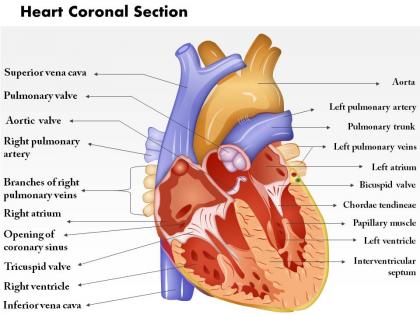
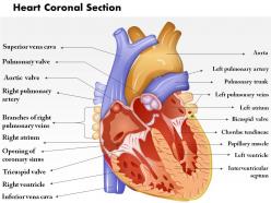
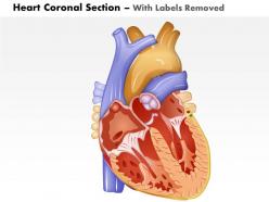
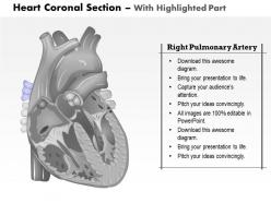
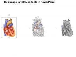
0514 heart coronal section medical images for powerpoint




Try Before you Buy Download Free Sample Product
 Impress Your
 Audience
 100%
100% Editable
 Save Hours
 of Time


Apply confidently with our 0514 Heart Coronal Section Medical Images For PowerPoint. You will get the go ahead.
People who downloaded this PowerPoint presentation also viewed the following :
0514 heart coronal section medical images for powerpoint with all 11 slides:
Open up your world with our 0514 Heart Coronal Section Medical Images For PowerPoint. Take a trip you will never forget.

FAQs for 0514 heart coronal section medical
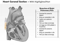
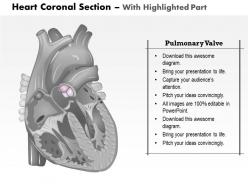
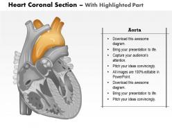
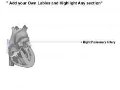
Coronal sections are perfect for seeing all four chambers spread out - atria on top, ventricles below. The septa between them show up really well too. Depending on where you cut, you'll catch the tricuspid and mitral valves, plus major vessels like the aorta and pulmonary trunk. Sometimes you get lucky and see coronary arteries if your slice hits just right. I honestly think coronal beats sagittal for learning this stuff - everything clicks spatially. Start with identifying the chamber walls first, then move to smaller details. Way less confusing than trying to piece things together from other angles.
Honestly, coronal sections are where it's at for cardiac imaging. They give you this perfect front-to-back view that shows chamber relationships way better than other planes. Wall thickness, septal defects, valve issues - it all just pops out at you. I always pull these up first when I'm looking at structural heart disease because the spatial stuff becomes obvious immediately. Sagittal cuts are fine I guess, but coronal views are clutch for complex congenital cases where you need to see how everything actually connects. Trust me, patterns that would take forever to figure out from axial images become clear instantly.
For what you're doing, MRI's gonna be your best bet - it's basically the gold standard for cardiac soft tissue imaging. You'll get amazing detail on myocardial structure and chamber anatomy, plus cine sequences show function really well. CT works too, especially for angiography, but there's that radiation thing to think about. Echo can technically do coronal views but honestly it's pretty limited compared to the other options. I mean, if you really need detailed coronal cardiac anatomy, cardiac MRI is what I'd reach for first. The image quality just blows everything else away for most of what you'll run into clinically.
So coronal sections are like front-to-back slices - imagine you're staring straight at the patient. Axial cuts? Those are your typical "loaf of bread" cross-sections from top to bottom. Sagittal splits things left-to-right, side view style. I'll be honest, coronal views mess with my head sometimes since we don't see them that often in cardiac stuff. But they're actually perfect for checking how the heart sits with everything around it. Super useful for surgical planning too. Next case you get, pull up all three planes and just mess around with them - that's how you'll really get the spatial thing down.
Dude, coronal sections are a game changer for cardiac surgery prep. They show you that front-to-back view that the other planes totally miss - like having GPS before cracking the chest open. Chamber relationships, valve positioning, vessel anatomy... it's all right there. Super helpful for spotting weird anatomical variants too (trust me, hearts can be quirky). Complex valve work or congenital cases? Don't even think about skipping these reconstructions. I learned this the hard way early in residency. Bottom line - always pull up those coronals before scrubbing in.
Dude, coronal heart sections are honestly your best friend for catching congenital stuff. That front-to-back view makes septal defects and valve problems way easier to spot than other angles. The four-chamber view especially - you can see both atria and ventricles at once, which is clutch for things like hypoplastic left heart or big VSDs. Great vessel relationships show up really well too. I always start with coronals when I'm looking at complex cases, though sometimes I get distracted by how clean the anatomy looks. Outflow tract issues are much clearer this way.
Honestly, the biggest issue is you're basically viewing the heart sideways, which makes chamber relationships way harder to read. Most cardiac pathology gets missed because coronal cuts go against the heart's natural anatomy instead of following it. Valve function assessment becomes tricky too. Also, let's be real - most cardiologists think in axial and sagittal planes, so coronal views just feel weird at first. The electrical conduction system? Forget about it, doesn't line up well with coronal at all. I'd definitely pair these with axial views, especially if you're dealing with anything complex. You'll get a much better picture that way.
Honestly, coronal sections are a game changer for cardiac pathology. You get this amazing cross-sectional view that shows wall thickness changes in cardiomyopathy way better than other planes. Regional wall motion problems from ischemic damage? Super obvious. Chamber dimensions are easier to measure too. I love using them for teaching - students actually get it when they can see the structure laid out like that. The anterior-to-posterior angle really clicks for understanding how different cardiac regions relate spatially. Try matching up your coronal views with clinical findings next time you're going through cases. Makes everything connect better.
Dude, digital enhancement is a game changer for heart coronal sections. Adjusting contrast and brightness brings out tissue differences you'd totally miss otherwise. Filters help reduce noise and sharpen those anatomical boundaries. False color mapping makes structures really pop too. Histogram equalization works great for dark regions - honestly saved my butt on a few tricky cases. Edge enhancement makes valve leaflets way clearer, same with septal defects. You should definitely experiment with different combinations on your next batch. The amount of hidden detail that shows up is pretty wild.
So you can stack those heart coronal images into 3D models that students actually walk through - imagine being inside the heart chambers, it's crazy cool. Most medical VR platforms take DICOM files directly, which saves you time converting stuff. Students can zoom into valves, rotate around vessel connections, even watch animated blood flow. Way better than staring at flat textbook pages, honestly. The spatial understanding hits different when you're virtually standing inside a beating heart. Oh and they can manipulate viewing angles however they want, so everyone learns at their own pace.
For heart coronal sections, start with ImageJ/FIJI - it's solid for basic stuff. DICOM viewing? Osirix or Horos work great. 3D Slicer is actually my go-to recommendation since it's free and handles most formats without being a pain. If you need fancy 3D reconstruction, Amira's good but pricey. Most cardiac labs use specialized tools like CardioVascular Toolbox or Circle CVI42, though honestly the learning curve is rough. I spent way too long figuring out CVI42 last year. Simple measurements? Basic DICOM viewers are fine. What are you planning to do with the sections? That'd help me point you toward the right tool.
Honestly, coronal sections are a game changer for coronary anatomy. You're basically slicing the heart front to back, so you can actually follow the left main as it splits into LAD and circumflex. Way better than trying to mentally reconstruct everything from axial slices - that's just painful. The right coronary shows up really nicely too as it wraps around. What I love is how clearly you can see the spatial relationships between vessels and chambers. Makes spotting anomalous origins so much easier. Definitely start with coronals next time you're looking at cardiac imaging - they'll orient you fast.
Dude, the image quality jumps are insane right now. 3T and 7T MRI machines are giving us coronal sections that make the old stuff look like potato quality - honestly kind of embarrassing how much we were missing before. AI reconstruction is helping a ton too, plus real-time cardiac imaging lets you actually see what's happening instead of guessing at motion artifacts. Oh, and the new contrast agents? Way better tissue differentiation. If you're stuck with ancient protocols, definitely bug your department about upgrading. The diagnostic difference isn't just nice to have anymore - it's actually changing patient outcomes.
Coronal sections are game-changers for mapping out the heart's conduction system. You'll get way better visualization of scar tissue and can actually trace those weird electrical pathways that cause problems. The septum and outflow tracts show up so much clearer than in axial views - honestly makes me wonder why some people still rely on those alone. Wall thickness measurements become super precise too. What's cool is you can basically plan your whole ablation strategy beforehand, figure out exactly where to target and what areas to steer clear of. Some docs even run simulations first, which seems a bit overkill but whatever works.
Definitely need to sort out the consent stuff first - make sure whoever donated these specimens actually agreed to educational use. You'll want clear policies about how students handle them respectfully, plus photo restrictions since these were real people. Some students might have religious or cultural issues with using human tissue, so maybe have backup options? I'd also be upfront about what the specimens are - don't try to hide that they're human. Honestly, the whole thing gets tricky fast if you don't have proper documentation. Set up respectful handling procedures and you should be good to go.
-
Helpful product design for delivering presentation.
-
The content is very helpful from business point of view.