

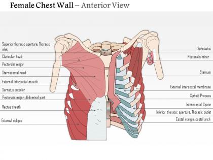
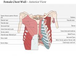
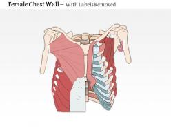
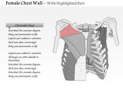
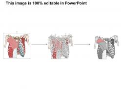
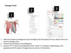
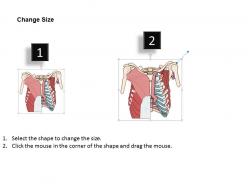
0514 female chest wall anterior view medical images for powerpoint




Try Before you Buy Download Free Sample Product
 Impress Your
 Audience
 100%
100% Editable
 Save Hours
 of Time


Indulge yourself with our 0514 Female Chest Wall Anterior View Medical Images For PowerPoint. Excite your senses with the variety we offer.
People who downloaded this PowerPoint presentation also viewed the following :
0514 female chest wall anterior view medical images for powerpoint with all 11 slides:
Our 0514 Female Chest Wall Anterior View Medical Images For PowerPoint are pretty cold blooded. They transfer all their heat to your ideas.
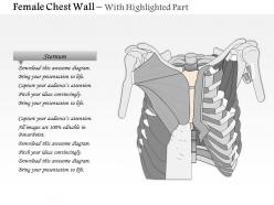
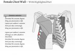
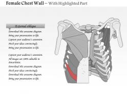
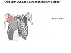

FAQs for 0514 female chest wall anterior view medical
So you're looking at the ribs, sternum, and clavicles mainly. The sternum runs down the middle - you've got the sternal notch at the top, then it goes into the manubrium and body. Ribs curve out from there, though honestly they're harder to count with breast tissue over the pec muscles. Clavicles make your upper border. Here's a tip that's actually useful - the nipple line usually marks the fourth intercostal space, which is super handy for clinical stuff. Counting ribs gets annoying but you can still see the basic framework underneath everything.
So breast density totally changes how your imaging looks. Dense tissue shows up white on mammograms and can hide stuff underneath - it's honestly super annoying sometimes. Fatty tissue is dark, so you get way better contrast. The difference between pre and post-menopausal patients? Night and day. Dense tissue also blocks more X-rays, so you might need to bump up your technique settings. CT and MRI help with the contrast between tissues, but dense areas still make things tricky to read. Just always think about density when you're planning and interpreting - it'll save you headaches later.
So when you're checking the chest wall, breast variations are totally normal - way more than you'd think. Size, shape, asymmetry, tuberous breasts, wide spacing - all fair game. Some people have accessory tissue along the milk line or even congenital absence like Poland syndrome. Age changes everything too, plus pregnancy and any surgeries they've had. I always document weird findings objectively but honestly? Function matters way more than how things look when you're deciding if something's actually clinically significant. The normal range is just massive.
So basically, as women get older their breast tissue changes a lot on imaging. Dense glandular tissue gets replaced with fat, which honestly makes mammograms so much clearer to read. The pectoralis muscle shrinks a bit too. You'll notice the skin gets thicker and breasts start to sag more - Cooper's ligaments stretch out over time. Oh, and the whole breast shape changes because of this. It's actually kind of ironic that older patients have better lesion detection since their tissue is less dense than younger women with really dense breasts.
So when you're examining the anterior chest wall, masses are obviously your biggest worry - both stuff you can feel and things that show up on scans. Look for any skin changes too, like dimpling or that orange peel texture. Nipple stuff matters - retraction, discharge, whatever looks off. I always check both sides because asymmetry is your best clue something's wrong. Don't miss the lymph nodes in the armpits either. Honestly, the systematic approach works best - just be thorough and compare everything bilaterally. Sometimes the subtle changes are what'll get you.
Hormonal changes really mess with breast tissue - you'll see major differences in density and appearance on chest imaging. Estrogen and progesterone make the mammary tissue expand and get way denser during puberty, pregnancy, and monthly cycles. Shows up as increased opacity. The areolar complex changes size and color too. Lactation makes it even more obvious - ductal prominence goes through the roof and volume shifts are huge. I always check where the patient is in their cycle because what looks concerning might just be normal hormonal stuff. Actually saves you from a lot of unnecessary worry calls.
Honestly, I'd go with ultrasound first - it's perfect for soft tissue stuff and you can see everything clearly: breast tissue, muscle, masses, fluid. Super quick too. If ultrasound doesn't give you the full picture, then MRI is great for those tricky cases where you need more detail. CT's solid if you're thinking bony issues or deeper pathology, though the radiation thing always makes me hesitate with younger patients. Mammography has its place obviously, but it's pretty limited for general chest wall assessment. I mean, ultrasound handles most of what you'll run into anyway.
So basically you're gonna see asymmetry and weird contours where they operated. Surgical clips and mesh show up as bright white spots - tissue expanders are super obvious once you know what you're looking for, they're these smooth round shapes. Watch for skin that looks thicker than normal and fluid collections from seromas. The architecture gets all wonky too. Here's the thing though - always compare both sides because that's when the subtle stuff jumps out at you. Oh and definitely check their surgical history so you don't miss any complications lurking around.
For breast masses and cysts, ultrasound is honestly your best bet. You can tell solid from cystic lesions super easily, check blood flow, and it's perfect when you're doing biopsies - the real-time guidance is clutch. No radiation either, which patients definitely appreciate, especially younger ones. It also works well for checking the chest wall muscles underneath if something seems off. The downside? It's really operator-dependent, and sometimes you'll miss deeper stuff. I always double-check findings with the physical exam though. If you need better soft tissue detail, MRI might be worth considering.
Knowing the chest wall anatomy makes breast cancer screening way more effective. You'll be able to tell what's normal tissue vs something suspicious. The pectoralis major insertion points are lifesavers - seriously, they prevent so many false alarms. Don't forget about the axillary tail either (that breast tissue extending toward the armpit). When you understand typical muscle boundaries and lymphatic patterns, distinguishing between benign variants and potential malignancies becomes much easier. Oh, and always use those anatomical landmarks to systematically hit all quadrants during exams. The tail of Spence gets missed a lot.
So basically when the chest wall gets messed up, it throws off how the whole breathing thing works. The rib cage can't expand right, so your lungs don't fill properly - tidal volumes drop and gas exchange gets wonky. Scoliosis and pectus excavatum are the usual suspects here. They create weird pressure imbalances that make you breathe all lopsided. Your diaphragm ends up doing way more work than it should. Honestly, spirometry tests are your best friend for catching this stuff early. Watch for people getting winded easily - that's when you'll really see how much their breathing mechanics are struggling.
Yeah, weight and exercise totally change how your chest wall looks. More body weight adds that layer of fat that smooths everything out - you won't see ribs or muscle definition as much. When you're leaner though, all those bony landmarks pop way more. Strength training makes a huge difference, especially if you're hitting pectorals and intercostals. Those exercises really bring out the muscle contours. Posture's probably the biggest game-changer honestly - slouching just ruins everything. Cardio doesn't directly change the chest wall shape but it'll shift your overall body composition over time. There's honestly such a wide range of what looks "normal" depending on all this stuff.
So look for the suprasternal notch first, then the clavicles and sternum - you know, the manubrium and body parts. Costal margins are key too. Nipple positions help but honestly they're all over the place depending on breast size. The midclavicular lines are probably your best friends for describing where stuff is located. Check if both sides look the same because when they don't, that's usually telling you something important. Inframammary folds matter too - I always forget those initially. Document everything relative to these landmarks so the radiologist knows exactly what you're talking about.
So the imaging tech has gotten crazy good lately for looking at chest wall stuff. Digital tomosynthesis basically slices through all that overlapping tissue that used to hide things on regular mammograms. MRI sequences are way sharper now too. There's this contrast-enhanced spectral mammography thing that's honestly pretty slick - it can tell dense tissue from actual lesions much better than before. The whole positioning process isn't as brutal either, which patients love. Bottom line? We're catching chest wall involvement way earlier and surgeons can plan with way more detail. Makes a huge difference compared to what we had just a few years back.
Netter's atlas is still my go-to after all these years - that book's just bulletproof. Grant's is solid too if you want another perspective. For digital stuff, Complete Anatomy and Visible Body are pretty sweet since you can actually rotate everything around and peel back layers. Khan Academy has decent anatomy modules (I know, sounds weird but they're actually good). Radiopaedia's great for real imaging examples. If you can swing it though, hands-on with cadaveric specimens or those plastinated models beats everything else. You really get the spatial relationships that way. Start with an atlas, then mess around with the interactive tools.
-
Excellent work done on template design and graphics.
-
Enough space for editing and adding your own content.