


0514 heart human anatomy medical images for powerpoint




Try Before you Buy Download Free Sample Product
 Impress Your
 Audience
 100%
100% Editable
 Save Hours
 of Time


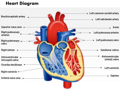
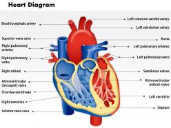
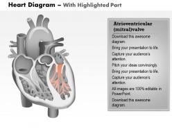
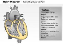
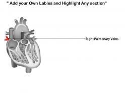
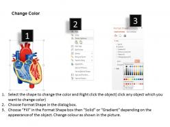
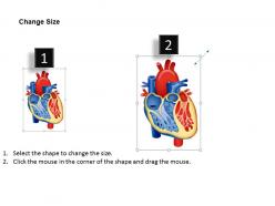
Whether you are a biology teacher or biology student, the available 0514 heart human anatomy medical images for PowerPoint is of much significance to your. This heart parts PPT PowerPoint presentation model has clearly laid down details related to the tiny little human organ known as heart. Make use of this human heart anatomy PPT presentation slide design to talk about your biology session where you need to share the detailing of the hear parts and functioning. This is a very detailed diagram with proper marking made for the heart. The available open heart PPT presentation visual is a flawless design in 3D presentation. The ease in use and application of the PowerPoint presentation visual ensures ease of download and customization. One simply needs to click on the download link provided below for human heart anatomy PPT presentation template and save it in desired format for using it in desired manner. Enhance goodwill with our 0514 Heart Human Anatomy Medical Images For PowerPoint. Benefit from a build up of friendly feelings.
People who downloaded this PowerPoint presentation also viewed the following :
0514 heart human anatomy medical images for powerpoint with all 11 slides:
Create a deadly combination with our 0514 Heart Human Anatomy Medical Images For PowerPoint. They will definitely click with your thoughts.


FAQs for 0514 heart human anatomy medical
So your heart's basically got four rooms - two up top (atria) that collect blood, two on bottom (ventricles) that pump it back out. Four valves keep everything flowing the right way, kinda like one-way gates. There's this wall called the septum down the middle that's super important - keeps the oxygenated and crappy blood separate. The whole thing sits in a protective sac too. Honestly, I always picture it like a tiny duplex with separate plumbing systems. Makes way more sense that way, at least to me!
Okay so all mammals basically have the same setup - two atriums, two ventricles, you know the deal. Size is where things get weird though. Horses have massive hearts because they're basically athletes, while tiny shrews have hearts beating at like 1000+ bpm which is honestly insane if you think about it. Most of the blood vessel stuff is pretty similar too, just positioned slightly different depending on the animal. Oh and smaller animals generally need way faster heart rates to keep up with their crazy metabolism. If you're studying this stuff, definitely focus on how heart size relates to what the animal actually does all day - that's where the cool patterns show up.
So basically your heart pumps blood to your whole body, right? But it also needs its own blood supply to keep working. That's where coronary arteries come in - they branch off the aorta and wrap around your heart, feeding it oxygen. Kind of weird when you think about it. Anyway, when these arteries get clogged up, parts of your heart muscle can literally die from lack of oxygen. That's a heart attack. All those procedures you hear about like stents? They're usually fixing blockages in these specific arteries. Your heart's pretty demanding honestly.
So your heart basically has this thing called the SA node - think of it as your body's natural pacemaker. It sends out electrical signals maybe 60-100 times a minute, which is wild when you think about it. The signals hit your atria first, making them squeeze, then go through the AV node to your ventricles. Pretty cool timing honestly - your atria need to fill up the ventricles before they pump everything out. Oh and if you ever notice your heart doing weird skips or racing? That's probably just the electrical stuff acting up, but definitely mention it to your doctor if it keeps happening.
So basically your heart's like two pumps working together. Right side gets the crappy deoxygenated blood from your body and sends it to your lungs. Left side takes that fresh oxygenated stuff back from the lungs. Here's the cool part - your left ventricle is way more jacked because it's gotta pump blood everywhere, not just to your lungs nearby. They never actually mix either, which is kinda wild when you think about it. Oh and if you're ever trying to explain this to someone, just say right = lungs, left = body. Works every time.
So basically, heart problems usually trace back to specific parts breaking down. Coronary arteries get clogged? That's your heart attacks and CAD right there. Valves start leaking or won't open properly - boom, valve disease. The electrical stuff goes haywire and you've got arrhythmias. Sometimes the muscle just gets weak or weirdly thick. I mean, it makes sense when you think about it - everything has a job. When I'm looking at patients, I try to figure out which piece isn't working. Makes the whole diagnostic process way less overwhelming, honestly.
So your heart has four valves that work like one-way doors - they let blood flow forward but slam shut to stop it from going backward. Pretty clever design, honestly. Without them working right, your heart would be fighting blood flowing the wrong way constantly, which makes it work way harder. It's like having turnstiles that don't work properly. When valves get damaged or don't close tight, you end up with problems like regurgitation or stenosis that really screw with how much blood your heart can actually pump out.
So when you're working out, your heart basically becomes a beast. Heart rate spikes, obviously, but the cool part is stroke volume - your heart chambers fill up more and squeeze way harder. Think of it like a balloon that fills bigger then contracts with serious force. Your cardiac output can jump 4-5x higher than when you're just chilling on the couch. Actually, next time you check your pulse mid-workout, you'll feel it's not just faster but way stronger too. Pretty wild how our bodies adapt honestly.
So basically the heart starts as this simple tube around day 18-19, then it folds and twists into the four chambers we know. First you get the primitive tube, then it loops to the right - and honestly, this part is make-or-break because if it goes wrong, that's where congenital defects come from. After that, the septa develop to separate everything into proper chambers. The foramen ovale stays open though, which is actually perfect for fetal circulation. Whole thing's done by week 10. When you're dealing with congenital heart cases, knowing this timeline really helps you figure out exactly when development went sideways.
Oh totally! So men usually have bigger hearts with thicker walls - women's are smaller but age way differently. Like, women get this concentric remodeling thing while guys get more eccentric changes (honestly the terminology is confusing but whatever). Aging screws with everyone though. Your heart walls thicken, valves get stiff from calcium buildup, and the electrical system slows down. It's actually super relevant when you're reading ECGs or echos. You can't just look at the results in a vacuum - gotta factor in whether it's a 70-year-old woman versus a 30-year-old guy.
So the big ones you'll run into are septal defects - basically holes between heart chambers. Patent ductus arteriosus is when this fetal blood vessel doesn't close like it should after birth. Coarctation means the aorta's too narrow. Tetralogy of Fallot sounds terrifying but it's actually just four defects grouped together, and docs have gotten really good at handling it. Valve problems like pulmonary or aortic stenosis pop up too. Oh, and most of these get spotted on prenatal ultrasounds now, so parents usually aren't totally blindsided anymore.
So the pericardium is basically this two-layer sac that wraps around your heart. There's fluid between the layers that cushions everything - honestly pretty smart design when you consider your heart's beating like 100,000 times a day. It keeps your heart from sliding around in your chest and maintains the right pressure for filling. Oh, and it prevents friction with each beat. When patients have pericarditis, that whole smooth system gets inflamed. That's why they get that sharp chest pain, especially when they breathe or move around.
So there are four main ones you'll deal with - echo (ultrasound), cardiac MRI, CT angiography, and cardiac cath. Echo's your go-to since it's cheap and non-invasive, shows real-time function pretty well. MRI gives crazy detailed soft tissue images and nails ejection fraction measurements. CT angiography rocks for seeing coronary arteries clearly. Cardiac cath is still the gold standard for finding blockages, but obviously it's more invasive - honestly kind of old school at this point but still necessary. I'd start patients with echo for baseline stuff, then get fancier based on findings.
So basically your heart is like this crazy efficient dual pump system. Right side sends blood to your lungs, left side pumps it everywhere else - at the same time. That left ventricle is super thick because it needs serious muscle to push blood through your entire body (we're talking thousands of miles of vessels, which is wild when you think about it). The valves are key though - they only open one way so blood can't flow backwards. Otherwise you'd just have blood sloshing around uselessly. Structure totally determines how well it works.
Your heart literally changes shape based on how you treat it. Smoking, junk food, being sedentary - all that stuff makes your heart walls thicker and forces it to work overtime. Regular cardio does the opposite though. It strengthens the muscle and makes everything more efficient. High blood pressure is the worst offender honestly, causing permanent structural damage. But here's the thing - you can still prevent more damage from happening. Start exercising, eat better, and you'll at least optimize what you're working with. Won't reverse everything, but it's definitely worth it.
-
Graphics are very appealing to eyes.
-
Top Quality presentations that are easily editable.