


0514 hip bone medical images for powerpoint




Try Before you Buy Download Free Sample Product
 Impress Your
 Audience
 100%
100% Editable
 Save Hours
 of Time


Cater for flaws with our 0514 Hip Bone Medical Images For Powerpoint. They cover up for any handicap.
People who downloaded this PowerPoint presentation also viewed the following :
0514 hip bone medical images for powerpoint with all 10 slides:
Our 0514 Hip Bone Medical Images For Powerpoint are fit for any function. They are empowering in every dimension.
-
0514 hip bone medical images for powerpoint
-
0514 hip bone medical images for powerpoint
-
0514 hip bone medical images for powerpoint
-
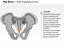
0514 hip bone medical images for powerpoint
-
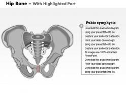
0514 hip bone medical images for powerpoint
-
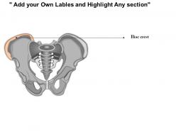
0514 hip bone medical images for powerpoint
-
0514 hip bone medical images for powerpoint
-

0514 hip bone medical images for powerpoint
-
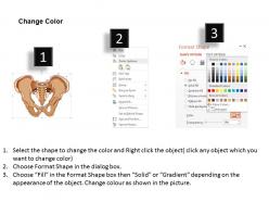
0514 hip bone medical images for powerpoint
-
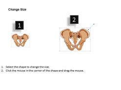
0514 hip bone medical images for powerpoint
FAQs for 0514 hip bone medical
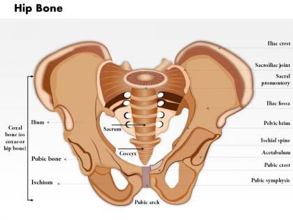
Okay so the hip bone breaks down into three parts - ilium (the big wing thing up top), ischium (what you're sitting on right now lol), and pubis at the front. They all meet up at this cup thing called the acetabulum where your thigh bone fits in. I swear anatomy naming is so weird sometimes. Start with the obvious stuff like iliac crest and that pointy anterior superior iliac spine. Oh and there's this big hole called the obturator foramen you can't miss. Once you get those main landmarks down, the rest isn't too bad.
So the 0514 hip bone is like your pelvis's main support system. Three bones fuse together there - ilium, ischium, and pubis - creating this ring that handles all the weight transfer from your spine to your legs. Pretty cool design if you ask me. Your major muscles attach there too, like glutes and hip flexors, which keep everything stable when you move. Oh, and here's the thing - when someone's having pelvic problems, I always look at that 0514 area first. Nine times out of ten, that's where you'll find what's throwing everything else off balance.
Fractures are huge, especially older people falling - that's like 90% of what you see. High-impact stuff causes dislocations, athletes get stress fractures from overdoing it. Hip dysplasia is when the socket forms weird, pretty common actually. Avascular necrosis is brutal - bone tissue just dies from bad blood flow. Happens after fractures or if someone's been on steroids too long. Pain complaints always make me think imaging first. X-rays usually, but sometimes you need MRI to catch the sneaky stuff.
So hip bones vary tons between people. Women have way wider pelvic inlets - like 90+ degree angles vs men's 70 degrees, all because of childbirth stuff. Men's bones are just more chunky overall with narrower cavities. Age makes a huge difference though - older folks get less dense bones, arthritis, sometimes full hip replacements. Kids are totally different since their growth plates don't fuse till late teens. I always forget how much this matters when looking at cases. What looks totally normal for a 70-year-old woman could be weird for a young guy.
Your hip bone is literally where all the action happens - connects your spine to your legs and takes a beating every time you move. The socket part (acetabulum) holds your thigh bone so you can rotate in basically any direction while staying stable. Crazy how much weight this thing handles daily! All your major muscles attach here too - glutes, hip flexors, the works. They're what give you power when you're moving and keep your legs positioned right. Oh and here's something cool - when your hip alignment is off, it messes with your knees and ankles too. Everything's connected down there.
So for hip imaging, docs usually start with regular X-rays - they're great for seeing bones, fractures, joint spacing, that kind of stuff. CT scans give you way more detail if there's a complicated break, but obviously more radiation. MRI's what you want for soft tissue problems like cartilage or ligament issues, plus it's really good for catching avascular necrosis (basically when bone tissue dies from poor blood supply - nasty stuff). Ultrasound works too, especially for kids since no radiation. Honestly, plain X-rays first unless there's reason to think it's soft tissue related.
So for hip bone problems, you've got a few surgical routes. Total hip replacement is pretty much the go-to - they swap out the whole joint with metal or ceramic parts. Hip resurfacing keeps more of your original bone, which is great if you're younger since you'll probably need another surgery later anyway. There's also osteotomy procedures that just realign everything without replacing the joint. Honestly, the choice depends on age, how active someone is, and what's actually wrong. I'd definitely get good imaging first though - and maybe send the tricky cases to ortho because some of this stuff gets complicated fast.
So basically your hip socket depth makes a huge difference for stability. Shallow sockets? You're gonna deal with impingement and wonky movement patterns. The whole thing is three bones fused together (ilium, ischium, pubis) that form this cup - and if the angle's off, you get uneven wear and pain. Think of it like a wonky ball bearing situation. Your walking, squats, everything gets thrown off. I'd honestly check your hip mobility pretty regularly. Oh and if you're feeling stiff or getting weird aches, maybe look into movement screening or something. Don't ignore that stuff.
Dude, your hip bone is seriously underrated - it's like the middleman transferring all the force between your upper and lower body when you move. Running, jumping, whatever you're doing, it takes a beating and spreads that stress through your pelvis so you don't get hurt. That socket joint (can't remember the fancy name) gives you crazy mobility while still being stable enough for all directions. Plus it's where your glutes and hip flexors attach, which is why those muscles matter so much. Honestly, I never thought about it until I tweaked mine last year. Just do some targeted hip exercises and you'll keep everything working right.
So crazy thing - your hip bone is actually three separate bones that fuse together when you're like 15-17. Ilium, ischium, and pubis just... merge. Wild, right? During childhood everything's growing fast and cartilage slowly hardens into bone. You hit peak bone density around 30, then lose about 1% yearly after that. Women get hit harder post-menopause because estrogen drops. The whole shape changes too - gets wider, joints wear down over time. Oh, and definitely get bone density scans after 50. That stuff's no joke.
Hip bones get absolutely wrecked by osteoporosis because they're constantly supporting your weight. The trabecular bone there loses density way faster than other types, which makes falls terrifying - especially around the femoral neck area. Hip fractures are honestly some of the worst outcomes I see. They often lead to major mobility loss and higher mortality rates, which sounds dramatic but it's true. Your patients really need bone density screenings and fall prevention plans. Early treatment with calcium, vitamin D, and meds can make a huge difference in keeping people independent. Prevention beats trying to fix things after a fracture happens.
So basically, working out makes your hip bones way stronger - weight-bearing stuff like walking actually tells your bones to build up more. Your glutes and hip flexors are like bodyguards for the joint, so when they're strong, there's less pressure on the bone. PT can fix wonky movement habits too (I had no idea I was walking weird until someone pointed it out). Your body's honestly incredible at adapting to movement. Just start easy with low-impact things and work up from there - don't go crazy right away.
Hmm, I don't recognize "0514 hip bone" as a standard medical term - sounds more like some kind of research code? Could be from a specific study or database you're working with. Without knowing where that designation comes from, I'm kinda shooting in the dark here for recent findings. Is this from a particular journal or clinical trial? Maybe your lab has its own classification system? Once you tell me the source, I can definitely help dig up whatever's new with that specific hip bone research. These research codes can be so random sometimes!
So your bones are basically tearing down and rebuilding constantly, which means you need the right fuel. Calcium and vitamin D are obvious ones, but protein matters too - plus magnesium and vitamin K (bone health is honestly way more complicated than I thought). Without enough of these, your hips just get weaker over time. That's how you end up with osteoporosis later. Actually track your calcium for like a week though - I did this and was getting maybe half of what I needed. Most people are walking around basically starving their bones without realizing it.
So CT and MRI scans are your best bet for getting those crazy detailed 3D views of bone structure. DEXA scans track density changes which is pretty useful. Honestly, 3D printing has been huge lately - you can literally print physical models to mess around with. There's also digital modeling software that lets you measure everything virtually (way easier than I thought it'd be). Oh, and advanced microscopy shows cellular details that used to be impossible to see. If you're doing morphological studies though, definitely start with high-resolution CT first.
-
Thanks for all your great templates they have saved me lots of time and accelerate my presentations. Great product, keep them up!
-
Innovative and attractive designs.