


0514 femur posterior view medical images for powerpoint




Try Before you Buy Download Free Sample Product
 Impress Your
 Audience
 100%
100% Editable
 Save Hours
 of Time


Stop putting off planning your presentation. With 0514 Femur Posterior View Medical Images For PowerPoint your design problems are solve in seconds.
People who downloaded this PowerPoint presentation also viewed the following :
0514 femur posterior view medical images for powerpoint with all 11 slides:
Encourage concerted action with our 0514 Femur Posterior View Medical Images For PowerPoint. The group will follow a common agenda.

FAQs for 0514 femur posterior view medical
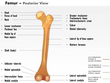
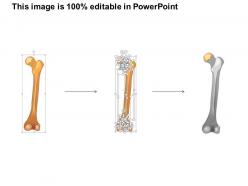
So the back of the femur has this big ridge called the linea aspera running straight down the middle - super easy to spot. Above it there's the gluteal tuberosity where your glutes connect (honestly such a weird name). At the bottom you'll see this smooth triangular bit called the popliteal surface where the ridge basically splits off. The medial and lateral condyles are there too, with that intercondylar fossa sitting between them. Oh and don't miss the adductor tubercle on the medial side. Pro tip: run your finger along all these landmarks while you study - it actually sticks way better than just staring at diagrams.
Honestly, that posterior view is clutch for understanding leg injuries. All the major muscle attachment sites are right there - you can spot where hamstrings and adductors connect at the linea aspera, which is exactly where athletes get those nasty avulsion injuries. The intercondylar fossa shows up clearly too, and that's where your ACL attaches so it helps explain the mechanics. Kids always seem to break their femurs in that supracondylar area, and it's way more obvious from behind. I always check the posterior view first now when I'm looking at any leg trauma - gives you the whole story pretty quickly.
So when you're looking at the back of the femur, focus on the linea aspera - that's where all the big thigh muscles attach. Hip pain patients? Check this ridge for muscle strain or weird bone stuff that's messing with their movement. The popliteal surface down by the knee is key too (I used to totally miss this one). You can feel some of these spots during your exam to match what you're seeing on imaging. Oh, and don't skip the posterior view on X-rays for fractures - you'll miss half the story otherwise. Honestly makes such a difference in understanding what's actually going wrong.
Honestly, imaging blows basic anatomy out of the water for seeing posterior femur stuff. CT scans are amazing for the linea aspera and where muscles attach - the detail is insane. MRI's your best bet for soft tissue around the posterior shaft. Sure, X-rays work for fractures, but they miss subtle posterior cortex problems sometimes which is annoying. 3D reconstructions totally change how you understand those weird posterior condyle shapes and the intercondylar notch. Start with multiplanar CT for crisp bone detail, then add MRI if you need the muscle correlation stuff.
Honestly, posterior femur variations can really mess with your surgical planning if you're not careful. The linea aspera prominence and gluteal tuberosity size vary way more than people think - I've seen cases where it completely changed the approach. Muscle attachment differences will affect your rehab protocols too. For hip replacements especially, those intercondylar notch dimensions matter for implant selection. Don't even get me started on popliteal surface variations - they'll screw up your safe zones around those vessels during posterior approaches. My advice? Always double-check your imaging beforehand, maybe grab 3D reconstructions for the tricky ones.
So the back of your femur is where all the action happens - there's this ridge called the linea aspera that's basically where your major leg muscles plug in. Your glutes, hamstrings, adductors all meet up there which is pretty genius for spreading out forces when you move. At the bottom you've got that notch where your ACL sits (weird to think about honestly). The whole setup lets you generate serious power for walking and running while keeping your leg stable. If you're studying this stuff, just follow those muscle attachment points along the ridge - it'll make the force directions way clearer.
So the linea aspera is that ridge running down the middle - start there, it's like your home base. Above it you've got the gluteal tuberosity where your glutes attach. The popliteal surface is this smooth triangular spot at the bottom, super clean looking. Honestly I always forget about those supracondylar lines but they branch off from the linea aspera too. Pro tip: just memorize the linea aspera first and everything else clicks. Way easier than trying to learn it all at once.
So the back of the femur is where tons of muscles attach. Gluteus maximus hooks onto the gluteal tuberosity, and adductor magnus grabs the linea aspera. Your hamstrings - biceps femoris, semitendinosus, semimembranosus - they're actually coming off the ischial tuberosity (which isn't technically femur but whatever). The linea aspera is honestly the main event though. That's where most adductors and hip extensors attach. If you're dealing with posterior thigh problems or thinking about surgery, you've gotta consider how messing with these attachment sites will mess with recovery. Those muscles don't like being disturbed.
So this view's pretty clutch for a bunch of stuff. Femoral neck fractures show up way clearer here - those Garden classifications that are basically invisible on AP. I've definitely missed posterior hip dislocations before checking this view properly, kinda embarrassing honestly. You'll catch femoral head AVN, SCFE in kids, plus any posterior acetabular breaks from trauma. The intercondylar notch is solid for knee pathology too. Always compare sides though - sometimes the only hint is weird asymmetry between hips.
Hey! So the pilaster (that ridge on the back of the femur) is way more prominent in European populations. African populations have totally different linea aspera patterns, and Asians often show unique popliteal surface shapes. Honestly, I find this stuff pretty cool when you start looking at the data. But here's the thing - these differences aren't just academic. They'll actually mess with your biomech analyses if you don't account for population ancestry. Same goes for forensic work. Don't make my mistake of ignoring population affinity early on!
So for posterior femur stuff, you've got two main options. Posterior hip approach works great for replacements and fracture repairs - gives you solid visualization of the posterior column and acetabulum. Just watch that sciatic nerve, it's honestly the trickiest part of the whole thing. Direct lateral might be better for shaft work though. The positioning can be a pain, but once you get your retraction down it's not too bad. I'd definitely brush up on the anatomy first if you haven't done many of these. Oh, and protect those posterior structures - learned that one the hard way during residency!
Dude, knowing the back of the femur is actually a game changer for PT work. Most people just ignore that whole area but that's where all the good muscle attachments are - biceps femoris, adductor magnus, all that stuff. Once you really get where everything inserts along the linea aspera, you can nail down exactly what's going wrong with someone's movement. Makes your strengthening programs so much more specific. The intercondylar notch thing affects knee mechanics too, which is huge for hamstring and adductor issues. I swear, start memorizing those posterior attachment sites and it'll totally change how you approach leg rehab. Worth the time investment for sure.
Dude, the femur's backside is like a timeline of how we learned to walk upright. That linea aspera ridge? It shows exactly how our muscle attachments changed when we ditched four-legged movement. Pretty cool stuff, honestly. The gluteal tuberosity got way more prominent to help us stand up straight, and you'll see similar changes in the adductor areas. Even the popliteal surface adapted for better knee stability - bipedalism required some serious bone remodeling. When you're comparing fossil hominids to modern humans and apes, these posterior features are where the evolutionary differences really pop out at you.
So the femur's posterior side is way more interesting than other long bones. There's this raised ridge called the linea aspera running down the center - that's where all your big muscles attach. Your glutes, hamstrings, adductors... they all need serious anchor points because the femur handles crazy forces. Other long bones? Pretty smooth on the back. But this ridge actually splits into two lines at both ends, making these triangular spaces. Pretty cool design if you ask me. Oh, and pro tip - if you're ever looking at bone specimens, that linea aspera is a dead giveaway you're holding a femur.
So basically, those posterior femur structures are where all your walking power comes from. The linea aspera is where your hamstrings and adductor magnus attach - super important for that push-off when you walk or run. Most people totally ignore the popliteal surface but it actually affects how your knee moves. When you're checking someone's gait, watch how their hip extension and stride length connect to these muscle attachment points. Oh, and try palpating along the linea aspera first - you'll immediately feel if there's weird tension or compensation happening. It's honestly one of the best starting points for gait analysis.
-
Innovative and Colorful designs.
-
Easily Editable.