


0514 pituitary gland medical images for powerpoint




Try Before you Buy Download Free Sample Product
 Impress Your
 Audience
 100%
100% Editable
 Save Hours
 of Time


Color up the event with our 0514 Pituitary Gland Medical Images For Powerpoint. They will create a brighter environment.
People who downloaded this PowerPoint presentation also viewed the following :
0514 pituitary gland medical images for powerpoint with all 11 slides:
Rejuvenate your team with our 0514 Pituitary Gland Medical Images For Powerpoint. Get them behind you with full sincerity.


FAQs for 0514 pituitary gland medical
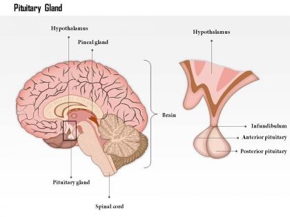
So your pituitary is like the boss of all your hormones - sits right under your brain and tells everything else what to do. The front part handles growth, reproduction, stress, thyroid stuff. Back part does water balance and labor contractions (wild, right?). It's seriously tiny but controls SO much. Your hypothalamus is basically its supervisor. Here's the thing though - any little problem with this gland can mess up your whole hormonal system. That's why even small changes on imaging can be a big deal. It's honestly amazing how much power something the size of a pea has over your entire body.
MRI is definitely your go-to here. The soft tissue contrast is amazing for that tiny pituitary area - you can spot microadenomas down to 2-3mm. Plus it shows if bigger tumors are squashing the optic chiasm or spreading into other stuff. Dynamic contrast studies are clutch because they help tell normal pituitary from tumor based on how they light up. CT honestly just sucks for this. Oh, and make sure you specifically ask for pituitary protocol with thin cuts and gadolinium - otherwise you might miss things.
So you'll mostly see adenomas - they're by far the most common. Size matters a ton here: micro ones are under 1cm, macro are above. Craniopharyngiomas and Rathke's cleft cysts show up pretty regularly too. Metastases pop up sometimes, along with lymphocytic hypophysitis and pituitary apoplexy, but those are way less common. Oh, and empty sella syndrome where the whole thing just looks... empty, which is honestly kind of wild when you first see it. Always check for optic chiasm compression and pay attention to those T1/T2 signal patterns - they'll tell you a lot.
When you've got hormonal issues, they usually show up as actual structural stuff on pituitary scans - adenomas, cysts, size changes that match the hormone chaos. Prolactinomas are everywhere and look like dark spots on T1 MRI. Too much growth hormone? Your sella turcica gets huge. Not enough hormones might give you an empty or half-empty sella instead. Here's the annoying part though - those tiny microadenomas under 10mm are seriously hard to catch, even with contrast. You really can't just look at pictures and call it good. Always match up your imaging with lab work and what the patient's actually feeling.
Definitely go with MRI and gadolinium contrast first - the soft tissue detail is incredible and you'll catch microadenomas down to like 2-3mm. Dynamic sequences are clutch because normal pituitary tissue enhances totally differently than tumors. CT's honestly pretty meh for sella stuff unless you're dealing with bigger masses or need to check for calcifications. Get high-res T1 and T2 in coronal and sagittal planes - that combo covers everything. Oh, and the dynamic imaging really makes a difference with enhancement patterns. Unless there's some reason they can't do MRI, start there. You'll miss way too much going straight to CT.
So basically it comes down to size - anything over 10mm is a macroadenoma, under 10mm is micro. The bigger ones are honestly way easier to catch because they're pushing stuff around, like displacing the optic chiasm or bulging out of the sella. Microadenomas just look like small dark spots in normal pituitary tissue. You'll usually see obvious mass effect with the large ones since they're literally changing the anatomy. Oh, and don't forget to check hormone levels too - that'll back up what you're seeing on the scan.
Ugh, honestly the tiny size is your biggest enemy here. Microadenomas under 10mm are impossible to spot sometimes. Then you've got all these normal pituitary structures that look sketchy but aren't actually pathology - super annoying. CSF pulsation screws up your images too, plus blood flow artifacts. The enhancement patterns are weird compared to surrounding tissue, so sequence timing matters way more than you'd think. Definitely use thin-slice T1 with gadolinium. But here's the thing - don't trust imaging alone if hormone levels are screaming something else. Clinical correlation saves your butt every time.
For pituitary tumors, MRI's definitely your go-to imaging. T1-weighted sequences show the anatomy really well, and T2 helps you tell tumor from normal tissue. The big thing is looking for mass effect - these tumors love squashing the optic chiasm above them, which gives patients that classic bitemporal hemianopia. They can also push into the hypothalamus or grow laterally into the cavernous sinus and mess with cranial nerves III through VI. Visual field defects are usually what makes patients come in first though. Oh, and don't forget to actually match what you're seeing on imaging with their symptoms - sometimes what looks scary on MRI isn't causing problems yet.
You literally can't see microadenomas without contrast - they're basically invisible on regular T1 since they look exactly like normal tissue. Once you add contrast though, boom, they show up as dark spots because they don't enhance like the rest of the pituitary does. Normal pituitary lights up pretty evenly with contrast, so anything that's too bright or too dark is immediately suspicious. Tumors, cysts, inflammation - they all have their own weird enhancement patterns that give them away. I've seen so many cases where the pre-contrast images looked totally normal, but post-contrast revealed obvious pathology. Don't even bother trying to read pituitaries without it.
Honestly, there's a pretty solid connection between what you see on imaging and the symptoms. Bigger adenomas usually mess with vision more because they're pressing on the optic chiasm. Hormone-secreting ones have their own patterns - prolactinomas will give you that classic galactorrhea/amenorrhea combo. Empty sella syndrome is interesting because the hormonal issues you see actually match up with how compressed the gland looks on MRI. Once you get the anatomy down, it all clicks. The trick is always checking your imaging against lab values though. Even if the gland looks totally normal but hormones are off, you've gotta dig deeper for microadenomas or infiltrative stuff.
MRI tech has gotten insanely good lately. We're seeing sub-millimeter detail now - stuff we'd completely miss even five years ago. The real game changer? Dynamic contrast imaging that shows blood flow in real time. Super helpful for catching those tricky functioning tumors that like to hide. 3T scanners are everywhere now (thank god, those old 1.5T images were terrible). Plus diffusion-weighted sequences help tell different tumor types apart. Bottom line - you can spot 2-3mm lesions confidently and actually plan surgery with crazy precision. It's honestly pretty amazing how far we've come.
Honestly, you can't wing pituitary surgery without solid imaging first. MRI sequences show you exactly where the tumor sits and how it relates to the optic chiasm and carotids. Coronal and sagittal views are your best friends for sellar anatomy - they're just perfect for this stuff. The images basically decide if you're going transsphenoidal or doing a craniotomy. Plus they'll warn you about nasty surprises like carotid encasement or suprasellar extension. I always sit down with radiology beforehand too. Four eyes are better than two when you're planning something this delicate.
So pituitary imaging is basically your GPS for endocrine stuff - you really can't manage these patients without it. MRI's the way to go (radiologists love their fancy contrast sequences but honestly they all blur together after a while). You'll spot adenomas, figure out size and invasion, see if they're actually pumping out hormones. The imaging tells you if someone needs surgery, meds, or just watching. But here's the thing - never go off images alone. Always match what you're seeing with hormone levels and what the patient's actually experiencing. That combo is what drives your whole treatment plan.
Yeah the pituitary is honestly a pain to image well. It's tiny - like pea-sized - and buried deep in this bony pocket called the sella turcica. Surrounded by all kinds of stuff that creates artifacts too. MRI works way better than CT for this since you get decent soft tissue contrast. I always go with sagittal T1-weighted sequences, they'll show you the clearest anatomy. Oh and definitely use thin slices if you're hunting for actual pathology - learned that one the hard way. The whole area's just cramped with the cavernous sinuses and optic chiasm right there.
So pituitary follow-ups are usually MRI at 3-6 months after surgery, then yearly for a few years. Honestly depends what type you're dealing with though. Adenomas need watching for regrowth, and functional tumors are trickier since hormones can get wonky. Post-radiation cases might need scans every 6 months initially - those changes happen super slowly which is kind of annoying. You'll want to stay tight with endocrine too since they're tracking hormone levels. The imaging plus labs together show if treatment's actually working.
-
Excellent Designs.
-
Visually stunning presentation, love the content.