


0514 female reproductive system medical images for powerpoint




Try Before you Buy Download Free Sample Product
 Impress Your
 Audience
 100%
100% Editable
 Save Hours
 of Time


Our 0514 Female Reproductive System Medical Images For PowerPoint deliver emphatically. They underline the strength of your conviction.
People who downloaded this PowerPoint presentation also viewed the following :
0514 female reproductive system medical images for powerpoint with all 11 slides:
Our 0514 Female Reproductive System Medical Images For PowerPoint are founded on facts. You can bet on their authenticity.
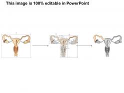

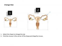
FAQs for 0514 female reproductive system medical
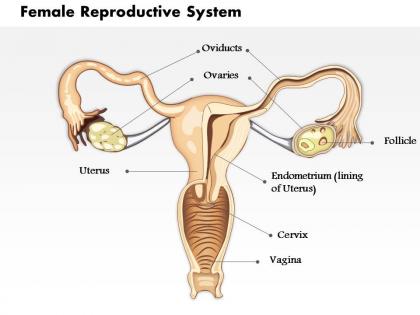
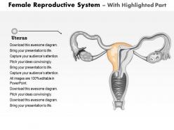
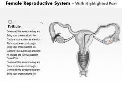
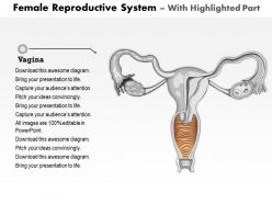
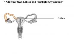
So you'll mainly be looking at ovaries, fallopian tubes, uterus, and vagina. Ovaries are those oval shapes on each side - that's where you spot follicles and any weird cysts or masses. The uterus is usually the biggest thing you'll see, especially that endometrial lining (thickness changes with her cycle). Fallopian tubes are honestly a pain to see unless something's wrong like hydrosalpinx. Oh and always check ovaries systematically when you're reviewing - endometrial thickness matters for like 90% of cases so don't skip measuring it.
So ultrasounds are perfect for this stuff because you get real-time images without any radiation. They'll show you exactly what's going on with cysts - size, location, whether it's just fluid or has weird solid bits mixed in. Same deal with fibroids. You can see how many there are and where they're sitting in the uterine wall, which doctors need to know before deciding treatment. The measurements are super precise too. Honestly, the whole thing is pretty cool how it distinguishes between different tissues. Plus you can do follow-ups to see if anything's changed over time.
MRI gives you insane soft tissue detail - perfect for fibroids, endometriosis, ovarian masses, that kind of stuff. No radiation either. CT's way faster though, which is clutch for emergencies like ovarian torsion. Plus it catches calcifications that MRI sometimes misses. Honestly? MRI can be total overkill for simple cases, but when you've got complex pelvic weirdness going on, nothing beats that tissue contrast. Oh and it's solid for cancer staging too. Most of the time I'd say start with ultrasound first, then jump to MRI if you need to dig deeper or can't figure out what you're looking at.
For endometriosis, they'll probably start with a transvaginal ultrasound since it's easier to get and catches most endometriomas pretty well. MRI is where it's at though - the detail you get is insane, especially for the deep stuff that ultrasound misses. That's what they use for surgical planning too. CT scans? Not really helpful unless they're checking for something else entirely. Honestly, most docs do the ultrasound first to see what's going on, then if you need surgery they'll order the MRI to map everything out properly. The newer MRI sequences are seriously impressive.
So basically, radiologists look for size, shape, wall thickness, and tissue density to spot problems. A healthy uterus looks pear-shaped with smooth walls and a thin endometrial line on scans. When something's off, you'll see weird shapes, masses, or thick walls. Fibroids show up as dark masses, adenomyosis makes the muscle look patchy - honestly, some of these findings can be pretty subtle at first. The malformation cases are usually more obvious since they totally change the normal shape. Takes time to get good at reading these! Always good to check measurements against normal ranges and see if symptoms match what you're seeing.
HSG catches tubal blockages really well, plus uterine stuff like fibroids and polyps. Structural problems show up too - bicornate uterus, septate uterus, that kind of thing. Adhesions from old infections or surgeries are pretty obvious once you get the hang of reading them - they create these weird filling defects. The contrast makes hydrosalpinx super clear when tubes are dilated with fluid. Oh, and cervical stenosis if you can't get contrast through at first (which honestly happens more than you'd think). Schedule between days 6-10 of her cycle for best results.
Dude, the depth perception alone makes such a difference - you can actually see how everything connects spatially instead of just guessing from 2D slices. Honestly, I've caught so many adhesions and structural weirdness that would've been easy to miss otherwise. Being able to rotate the images around is clutch too. The way the ovaries, tubes, and uterus relate to each other becomes super obvious once you see it in 3D. I know it sounds like overkill sometimes, but pull up those reconstructions next to your standard views on your next tricky case. Trust me, it'll click differently when you can visualize the actual spatial relationships instead of trying to piece together fragments.
Honestly, the anatomy down there is just complicated as hell. Every patient's different too, which makes spotting what's actually abnormal pretty tough. Image quality is another headache - you get bowel gas blocking everything or larger patients where you can't see much. Timing matters with the menstrual cycle since hormones change how things look. I'd say always check the clinical notes first, helps give context. And if something seems off but you're not sure? Just recommend another scan later. Better safe than sorry with pelvic imaging.
Hey! So imaging tech has totally changed the game for diagnosing reproductive stuff. We're spotting issues way earlier now. High-res ultrasounds show crazy detail - like you can see ovarian cysts, fibroids, even how thick the endometrial lining is. MRI is incredible for tricky cases (seriously, the image quality still amazes me). 3D ultrasound catches uterine problems that regular 2D would miss completely. Oh, and patients love that it's way less invasive than the old methods. Doctors can make diagnoses faster and feel way more confident about what they're seeing.
Okay so the big thing is consent - these are real people who trusted their doctors, you know? Make sure patients actually agreed to educational use when the photos were taken. Strip out anything that could identify them too. Honestly, reproductive imagery can be pretty intense for some people, so definitely give a heads up beforehand. Cultural stuff matters here - what's clinical to you might be shocking to others. I'd stick with reputable medical sources that did their homework on consent protocols. This kind of thing can blow up legally if you're not careful about it.
Imaging is huge for pregnancy monitoring - think of it as your window into what's happening with baby and mom. Ultrasounds track growth, check organs, and monitor fluid levels plus placental position. They're totally safe to repeat, which is why you get so many throughout pregnancy. Honestly, the technology still amazes me sometimes. If there's something concerning, MRI can dig deeper into placental issues or fetal abnormalities that ultrasound might miss. The whole point is catching problems early rather than waiting for symptoms to show up. Regular scans help doctors make better care decisions before things get complicated.
Ugh, yeah - regular imaging is pretty limited for reproductive stuff. Ultrasounds miss tiny lesions and don't show soft tissue well. X-rays? Basically useless for ovaries or uterus. MRI works great but costs a fortune and takes forever - insurance companies hate approving it unless you're already dying lol. CT scans mean radiation exposure, which isn't ideal when you're trying to have kids someday. The real problem is catching things early, like endometriosis or small cysts. Honestly, doctors usually end up combining different imaging methods because no single one gives the full picture.
You can't wing gynecologic cancer surgery without good imaging first. MRI gives you the clearest picture of tumor spread and lymph nodes - way better soft tissue detail than anything else. CT's solid for catching distant mets you might miss. PET scans are honestly clutch for finding hidden disease. Before any radical hysterectomy or debulking, I need to know exactly what I'm walking into. Can't just order a basic "pelvic MRI" though - you've got to specify the right sequences or you'll get garbage images that don't help anyone. Makes all the difference in surgical planning.
Oh man, imaging is everything in fertility work! You're basically flying blind without it. Transvaginal ultrasounds are your best friend - tracking follicles, measuring that endometrial lining, guiding egg retrievals. HSGs are kind of awful but show if tubes are blocked (which is huge). MRIs catch structural problems like fibroids that mess with implantation. The tricky part? Timing matters so much with cycle phases to get accurate reads. I've seen docs miss stuff just because they imaged at the wrong time. Real-time guidance during procedures is a game-changer too.
Honestly, just taking a few minutes to explain imaging makes such a huge difference. Patients freak out way less when they know what an ultrasound will actually show them. They're more likely to show up for follow-ups too, which - let's be real - half the battle right there. I've caught so many ovarian cysts early just because women understood why we needed that second scan. When patients get what they're looking at in results, they ask way better questions. Sometimes they'll even spot stuff we might've missed on first glance. It sounds obvious but most docs skip this step completely.
-
Great quality slides in rapid time.
-
Excellent work done on template design and graphics.