


0514 human heart medical images for powerpoint 2




Try Before you Buy Download Free Sample Product
 Impress Your
 Audience
 100%
100% Editable
 Save Hours
 of Time


Our 0514 Human Heart Medical Images For PowerPoint 2 are a definite guarantee. They fulfill on every account.
People who downloaded this PowerPoint presentation also viewed the following :
0514 human heart medical images for powerpoint 2 with all 10 slides:
Have it all. Our 0514 Human Heart Medical Images For PowerPoint 2 are high quality affordable and ready at a moment's notice.
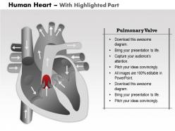
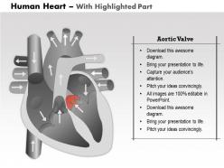
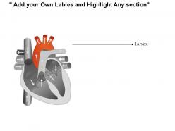
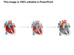
FAQs for 0514 human heart medical images
So the main ones are echo, CT angiography, cardiac MRI, and nuclear stuff like SPECT or PET. Echo's probably what you'll use most - it's quick, non-invasive, great for valves and ejection fraction. CT angiography shows amazing 3D coronary views (seriously, the resolution now is insane). MRI's your best bet for precise tissue characterization and chamber measurements. Nuclear imaging checks blood flow and metabolism - though honestly it's not my favorite, takes forever. Really depends what you're trying to figure out though. What's the clinical question you're dealing with? That'll help narrow down which one makes sense.
Okay so echo is like watching your heart live with ultrasound - super quick, maybe 20 minutes, and you can do it right at the bedside. MRI though? That's the fancy stuff with magnetic fields, takes like an hour plus but holy crap the detail is insane. You'll see tissue damage, scarring, inflammation that echo totally misses. The thing is, echo's perfect for valve problems since it's moving in real-time. MRI gives you those gorgeous static shots with crazy resolution. Honestly I'd go echo first for most patients - it's cheaper and faster. Save MRI for when you really need to dig deeper into tissue issues.
Dude, CT scans are crazy good for heart stuff now. You can get calcium scores to see plaque buildup, plus the angiography shows exactly where blockages are. Image quality has gotten so much better - like, night and day difference from a few years back. For chest pain patients, it's honestly become my go-to instead of jumping straight to invasive procedures. You'll catch disease early and figure out how bad it actually is. I mean, the detail is pretty incredible when you see those 3D reconstructions. Definitely worth considering as your first imaging choice for anyone with cardiac risk factors.
Ok so basically you're hunting for blockages and narrowed spots in the coronary arteries. The contrast dye lights up the vessels - any dark areas or super thin sections? Those are your problem spots. Left main artery blockages are scary stuff, so always check that first. Then work through each major vessel systematically. Collateral circulation is cool to spot too - shows how the heart creates workarounds when arteries get blocked. Document stenosis percentages as you go. Honestly takes practice but you'll get faster at pattern recognition. My attending always said think like a plumber looking for clogs.
Dude, the heart imaging stuff they're doing now is honestly incredible. Machine learning can spot things in CT scans and MRIs that doctors might totally miss - and way faster too. There's this 4D imaging that shows your heart beating in real time, which is kinda mind-blowing if you ask me. Better contrast agents make everything clearer now. The cool part? They catch problems super early, so people get treated before things get really bad. Makes such a huge difference in how patients do long-term.
Ultrasound is perfect for heart stuff - you get real-time images without radiation, which is clutch for bedside checks. ICU docs love how portable it is. But here's the thing: image quality tanks with heavier patients or anyone with lung issues since sound waves can't push through air or thick tissue well. You're also totally screwed if the operator sucks at it. Patient has to be positioned just right too. Honestly, for anything complex you'll probably need CT or MRI backup anyway. It's solid for checking function and initial screening though. Just don't expect miracles with tricky cases.
So MRI and echo are your best options - literally zero radiation. CT angiography hits you with about 1-15 mSv depending on the machine, and nuclear stress tests can go 9-25 mSv which is honestly pretty high. The newer CT scanners are way better though, radiation's dropped a ton. Chest X-rays are basically nothing at 0.1 mSv but you won't see much detail. I'd definitely think about radiation exposure when picking tests, especially if you're young or might need follow-ups later. Some of these numbers add up fast if you're not careful.
So cardiac MRI can pick up a bunch of stuff - cardiomyopathies, heart attacks, congenital defects, valve problems, pericardial issues. The detail you get is honestly incredible compared to echo sometimes. They also use it for ARVC, cardiac tumors, checking if heart muscle's still viable after MIs. Oh and the T1/T2 mapping thing is really good for spotting inflammation and scarring. If you need to figure out whether someone's got ischemic vs non-ischemic cardiomyopathy, MRI with gadolinium contrast is pretty much the gold standard for nailing that diagnosis.
Honestly, 3D imaging is so much better than regular echo. Instead of flat slices, you get the whole spatial picture - can actually see how chambers connect and watch blood flow in real time. The cool part is you can rotate everything around, zoom in on weird spots, and catch structural problems that 2D views totally miss. Volume measurements are way more accurate too, plus you can track wall motion through the entire cycle. I always forget how much clearer things become until I'm looking at a tricky case. Definitely ask for the 3D reconstruction next time - it'll save you from that headache of piecing fragments together.
Color Doppler is a game-changer - you can literally watch blood flow in real-time and see exactly which direction it's going. Red flows toward your probe, blue flows away, which makes spotting valve problems super obvious. I swear it's like having x-ray vision when you're checking cardiac function! You'll catch regurgitation and stenosis right away just from those color patterns. Plus the velocity readings let you calculate pressure gradients across valves. Oh, and don't forget to tweak your angle and gain settings - I've missed subtle flow issues before because my settings were off. Those little details can totally change what you're seeing.
Dude, you absolutely need those scans before any heart surgery. CT and MRI show you everything - where the vessels are, what's abnormal, exact measurements. It's honestly like having superpowers or something. You can map out the safest way in and catch problems before they blindside you during the actual procedure. Cuts down your operative time too since you're not figuring things out as you go. Trust me, spend the time reviewing those images in your pre-op sessions. Way better than getting surprised with your hands already inside someone's chest.
Cardiac imaging with AI is honestly getting wild right now. Deep learning can spot coronary artery disease from CT scans automatically, and some models predict heart attacks way better than old-school methods. The coolest part? Surgeons get real-time feedback during actual procedures - like having a super smart assistant right there. Oh, and federated learning is huge too - hospitals can collaborate on AI training without sharing patient data, which is smart given all the privacy stuff. If you're looking to implement this, honestly just start with whatever imaging workflow eats up most of your time. That's where you'll actually see results fast.
Honestly, imaging and symptoms usually match up pretty well, but you'll get some curveballs. I've seen patients with terrible stenosis still running around fine, while others with minor stuff can barely function - bodies are weird like that. Most of the time though, low ejection fraction means they're tired and short of breath. Wall motion problems? Usually chest pain. Bad valve function typically shows up as poor exercise tolerance. The imaging basically explains what they're feeling - gives you the whole picture instead of just guessing. Just don't look at scans without talking to your patient first.
Look, the big ones are consent, privacy, and who actually gets access. Advanced heart imaging picks up way more than patients realize - genetic stuff, random findings they weren't expecting. So your consent forms better spell out exactly what you're collecting. Data security's huge too since these scans create massive amounts of sensitive info that hackers would love. Oh, and honestly? The cost thing pisses me off - these technologies are expensive so you'll probably see richer areas getting better access while everyone else gets left behind. Just make sure people know what they're signing up for and how their data gets stored.
Dude, the imaging quality makes such a huge difference. You can actually see where all the blockages are instead of guessing, plus you'll know the exact vessel sizes before you start. Any weird anatomy shows up clearly too - saves you from those "oh shit" moments mid-procedure. I used to hate working with those fuzzy old images. Now you can pick the right equipment ahead of time and your whole approach is way more strategic. Honestly cuts down on complications and you're not fumbling around trying to figure out what's going on. Check how your next case's imaging matches what you find.
-
Perfect template with attractive color combination.
-
Perfect template with attractive color combination.