

0614 simple cuboidal epithelium medical images for powerpoint




Try Before you Buy Download Free Sample Product
 Impress Your
 Audience
 100%
100% Editable
 Save Hours
 of Time


Our 0614 Simple Cuboidal Epithelium Medical Images For Powerpoint can be the bane of dogma. They discourage any domineering attitude.
People who downloaded this PowerPoint presentation also viewed the following :
0614 simple cuboidal epithelium medical images for powerpoint with all 8 slides:
Get them in the groove with our 0614 Simple Cuboidal Epithelium Medical Images For Powerpoint. Encourage them to follow given directions.


FAQs for 0614 simple cuboidal epithelium medical
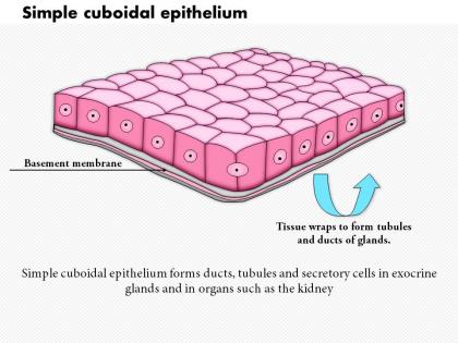
So these cells are basically built for moving stuff around - secretion and absorption is their whole thing. You'll spot them in kidney tubules doing all that filtering work, plus they make up the secretory parts of glands like your thyroid. The cube shape is actually perfect for this since there's room for all the organelles they need. Honestly, it's pretty cool how form follows function here. When you're looking at slides, single layers of cubes usually mean some heavy biochemical lifting is happening. Oh, and salivary glands too - almost forgot those.
So cuboidal cells are basically built like little workhorses - the cube shape gives them way more internal space than those flat squamous cells. More room means they can pack in tons of mitochondria and ER to do all that secretion work. They stack together nicely too, which is honestly pretty efficient. Short height keeps things moving quickly between surfaces, but there's still enough space for all the cellular machinery they need. You'll spot these in kidney tubules and gland ducts where they're constantly filtering and secreting stuff. Form follows function, you know? It's actually kind of cool how perfectly designed they are for the job.
So you're gonna find simple cuboidal epithelium mostly in kidney tubules, thyroid glands, and those smaller glands like salivary and sweat glands. Pancreas and liver ducts too. Basically these cube-shaped cells are workhorses - perfect for secretion and absorption since they're compact but still pack enough cytoplasm for all that cellular machinery. Pretty smart design if you ask me. When you're looking at slides, just hunt for those distinctive cube shapes with nuclei right in the center. That's your telltale sign you've found it.
So basically, cuboidal cells look exactly like their name suggests - little cubes! They're as wide as they are tall, unlike squamous cells that are super flat or columnar ones that are tall and skinny. Under the microscope they're pretty distinctive once you get the hang of it. You'll see them mostly in kidney tubules and gland ducts where absorption and secretion happen. Honestly, I always just look at the height-to-width ratio first - it's like the cheat code for identifying them. Squamous is more for protection, columnar does the heavy lifting in your intestines. Pretty straightforward compared to some of the other tissue types we've had to memorize!
So kidney tubules are lined with simple cuboidal epithelial cells - they're like the workhorses doing all the heavy lifting. These cube-shaped cells are actually perfect for the job because they can efficiently move stuff between your blood and the filtrate. You'll see them in the proximal and distal convoluted tubules, actively grabbing sodium, glucose, amino acids and shuttling them back to your bloodstream. At the same time they're dumping waste and extra ions into urine. Honestly, it's wild how such basic-looking cells handle so much transport work. Focus on that structure-function relationship when you're studying nephrons!
So basically microvilli are like tiny fingers that stick out from cuboidal epithelial cells. They massively increase surface area, which is perfect for absorption and secretion. You see them a lot in kidney tubules - makes sense since kidneys need to filter like crazy. More surface area = more room for all those transport proteins and channels to do their thing. It's honestly pretty clever how the body maximizes efficiency in such small spaces. Glandular tissues use this trick too when they need serious substance exchange happening fast.
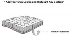
So the basement membrane is like the foundation that keeps those cuboidal epithelial cells stuck in place - without it they'd just drift off. It's also this selective barrier that controls what moves between the epithelium and the tissue underneath. Honestly, I always think of it as a bouncer deciding who gets in lol. Plus it helps maintain cell polarity and sends chemical signals to keep everything functioning right. Oh and when you're examining samples, definitely check if the basement membrane looks intact because if it's damaged, the whole epithelium is probably messed up too.
So simple cuboidal epithelium is basically what keeps endocrine glands running smoothly. These cube-shaped cells line stuff like thyroid follicles and pancreatic islets. They're pretty smart - they can ramp hormone release up or down depending on what your body needs at the moment. They also handle ion transport and keep fluid levels balanced inside the glands, which honestly seems like a lot for such tiny cells! Before hormones get released, these cells can even modify them first. Oh and if you're looking at endocrine diseases, definitely check if the cuboidal cells are messing up - that usually throws off the whole feedback system.
Oh right, so those cuboidal cells get trashed by a bunch of stuff. Diabetes really screws them up - hits the kidney tubules hard. You'll also see damage from toxins or when blood flow gets cut off (acute tubular necrosis is nasty). Chemical exposure does it too, plus nephrotoxic meds are basically poison to these cells. Honestly, infections and chronic inflammation just slowly chip away at them over time. When you spot this kind of damage, think metabolic problems first, then toxic exposure, or circulation issues. Those are your usual suspects anyway.
So basically, simple cuboidal epithelium bounces back pretty well when it gets damaged - the cells multiply fast and regenerate nicely. The basement membrane usually stays put, which is honestly clutch because it gives new cells something to build on. Now here's where it gets interesting though. If there's chronic irritation or inflammation, these cells can actually transform into completely different cell types - it's called metaplasia. You see this a lot in kidney tubules after toxin exposure. They can also get bigger or smaller depending on what's demanded of them functionally. Quick tip: watch for changes in cell height and where the nuclei sit when you're looking at samples. That's your first clue something's going wrong.
For cuboidal epithelium, CK7 and CK19 are your go-to cytokeratins - they're basically the gold standard. E-cadherin works great too since it shows those tight junctions between cells. EpCAM is decent for general epithelial stuff, though I honestly think cytokeratins are way more reliable. Actually, if you're doing multiple samples, I'd just do a CK7/CK19 double stain from the start. Saves you the hassle of running separate stains later. They're super consistent across different tissues too, which is nice when you're not sure what you're dealing with.
So simple cuboidal epithelium is basically the middle ground for healing - not crazy fast like simple squamous but way better than the layered stuff. These cells can still divide pretty well, especially in glands and kidney tubules. Since it's just one layer, replacement isn't super complicated, but here's the thing - they do specialized work like secretion so it takes longer to get back to full function. I'd say expect decent regeneration, maybe a few days to weeks depending where we're talking about. Not lightning speed but solid recovery overall.
So basically, when those cuboidal cells in your kidneys, thyroid, and pancreas start getting messy - like losing their organized shape or multiplying too fast - that's when pathologists get worried. It's usually one of the first signs something's going wrong cancer-wise. Renal cell carcinoma does this thing where it completely trashes the normal cell architecture, which is pretty obvious under a microscope. The whole epithelial-to-mesenchymal transition stuff is getting really big in research right now. Honestly sounds fancy but it's just cells changing their behavior. Definitely worth following those studies since they're becoming solid biomarkers for catching cancer early.
So the cuboidal shape is actually perfect for this stuff - you get good surface area but it's still thin enough for quick transport. Kidney tubules are a great example of this in action. The cells pack super tight together which creates these selective barriers that can control what passes through. Honestly, it's kind of genius how they balance being sturdy for protection while staying efficient for absorption and secretion. When you're looking at samples, that orderly cube pattern is usually a dead giveaway about function. Just make sure you check where it's located too - that'll tell you if it's mainly secreting or absorbing materials.
Confocal microscopy is probably your best bet for live cuboidal epithelium - good resolution and pretty straightforward to use. Two-photon is amazing if you can get access since it goes deeper without cooking your samples (trust me on this one). OCT gives you nice structural overviews but the resolution's kinda meh. You could also try live-cell fluorescence if you're tracking specific proteins or watching cell behavior. Honestly, I'd just start with confocal though - covers most bases and won't make you want to pull your hair out learning it.
-
Awesome use of colors and designs in product templates.
-
Excellent work done on template design and graphics.