

0714 t tubule and sr terminal cisternae associations medical images for powerpoint




Try Before you Buy Download Free Sample Product
 Impress Your
 Audience
 100%
100% Editable
 Save Hours
 of Time


Convince them it's not hard with our 0714 T Tubule And SR Terminal Cisternae Associations Medical Images For Powerpoint. Your argument becomes easier to believe.
People who downloaded this PowerPoint presentation also viewed the following :
0714 t tubule and sr terminal cisternae associations medical images for powerpoint with all 9 slides:
If you have to create many presentations with that revolve around the same theme our custom created 0714 T Tubule And SR Terminal Cisternae Associations Medical Images For Powerpoint can be a perfect fit. This will ensure you don't have to create a new presentation each time you need to make an update.

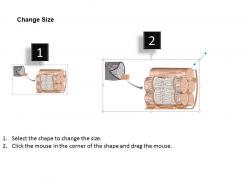
FAQs for 0714 t tubule and sr terminal cisternae associations medical
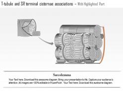
T-tubules enable action potentials to penetrate deep into muscle fibers, ensuring synchronized calcium release from the sarcoplasmic reticulum across the entire fiber diameter. Through this strategic positioning, T-tubules facilitate rapid, coordinated muscle contraction by transmitting electrical signals to SR terminal cisternae, ultimately delivering uniform calcium availability and efficient excitation-contraction coupling throughout the muscle fiber.
T-tubules feature narrow diameter, extensive branching networks, and strategic positioning at sarcomere boundaries, enabling rapid electrical signal transmission deep into muscle fibers. These structural characteristics streamline excitation-contraction coupling by ensuring synchronized calcium release across the entire fiber cross-section, with cardiac and skeletal muscle systems finding that this architecture delivers coordinated contraction and enhanced contractile efficiency.
T-tubule and SR interactions center on calcium-induced calcium release, where T-tubule depolarization activates dihydropyridine receptors that trigger ryanodine receptors in SR terminal cisternae. This coupling mechanism enables rapid, synchronized calcium release across muscle fibers, with cardiac and skeletal muscles utilizing slightly different receptor configurations, ultimately delivering the precise calcium concentrations needed for efficient contraction and enhanced muscular performance.
The morphological arrangement of terminal cisternae enhances calcium storage through enlarged sarcoplasmic reticulum regions positioned strategically at sarcomere boundaries, creating high-capacity reservoirs with increased surface area for calcium-ATPase pumps. This structural organization enables rapid calcium release via clustered ryanodine receptors, while proximity to T-tubules ensures synchronized excitation-contraction coupling, ultimately delivering faster muscle contraction and more efficient relaxation cycles.
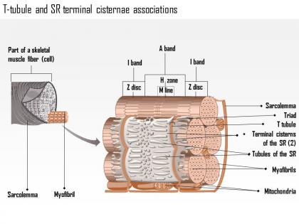
Skeletal muscle features precise triads where T-tubules associate with two SR terminal cisternae at Z-line junctions, while cardiac muscle displays diads with T-tubules connecting to single SR cisternae at Z-lines. These structural differences enable skeletal muscle to deliver rapid, synchronized calcium release for powerful contractions, whereas cardiac muscle provides more regulated calcium handling for sustained, rhythmic contractions, ultimately supporting their distinct functional demands in movement versus circulation.
Junctional feet, which are ryanodine receptors in terminal cisternae, dramatically enhance calcium signaling by creating direct communication channels between T-tubules and sarcoplasmic reticulum during muscle contraction. These protein structures enable rapid, synchronized calcium release across muscle fibers, streamlining excitation-contraction coupling and ultimately delivering faster, more coordinated muscular responses essential for efficient physiological performance.
Calcium-induced calcium release (CICR) represents the primary signaling pathway, where voltage-gated L-type calcium channels in T-tubules trigger ryanodine receptors in the SR terminal cisternae. This excitation-contraction coupling mechanism enables rapid, synchronized calcium release throughout muscle fibers, with cardiac and skeletal muscle systems finding that this strategic association delivers enhanced contractile efficiency and precise temporal control of muscle activation.
T-tubule density directly enhances force generation by enabling more synchronized calcium release across muscle fibers, ensuring uniform contraction throughout the fiber's cross-section, and minimizing delays in excitation-contraction coupling. Higher densities streamline calcium delivery to contractile proteins, with cardiac and skeletal muscles demonstrating that increased T-tubule networks ultimately deliver greater contractile efficiency and stronger, more coordinated force production.
T-tubule and SR dysfunctions significantly compromise muscle performance by disrupting calcium handling, excitation-contraction coupling, and membrane integrity, leading to weakened contractions, delayed relaxation, and cellular damage. These impairments manifest in conditions like muscular dystrophy, heart failure, and myopathies, where compromised calcium release and reuptake ultimately result in reduced muscle strength, increased fatigue, and progressive tissue degeneration across cardiac and skeletal muscle systems.
During muscle development, T-tubule and SR associations undergo progressive maturation from sparse, irregular connections in embryonic stages to highly organized dyadic and triadic junctions in mature muscle. This developmental progression enhances calcium handling efficiency, contractile force generation, and excitation-contraction coupling precision, with cardiac and skeletal muscle tissues ultimately achieving optimal performance through these increasingly sophisticated membrane associations.
Electron microscopy, confocal fluorescence microscopy, super-resolution imaging, immunohistochemistry, and calcium indicator dyes are commonly used to visualize T-tubule and SR structures in muscle tissue. These advanced imaging techniques enable researchers to examine structural relationships, calcium dynamics, and protein distributions with high resolution, while fluorescent labeling and specialized dyes facilitate real-time monitoring of muscle contraction mechanisms, ultimately delivering enhanced understanding of muscle physiology and disease states.
Ion channels in T-tubule and SR complexes include voltage-gated calcium channels, ryanodine receptors, potassium channels, and sodium-calcium exchangers, creating sophisticated communication networks for muscle contraction. These channels work together by enabling rapid signal transmission, facilitating calcium release and reuptake, and maintaining ionic gradients, ultimately delivering precise contractile control and enhanced muscle performance across cardiac and skeletal muscle systems.
Calcium-induced calcium release creates a critical amplification mechanism where initial calcium entry through T-tubule voltage-gated channels triggers massive calcium release from SR terminal cisternae through ryanodine receptors. This process enables cardiac and skeletal muscle cells to achieve the substantial calcium concentrations needed for effective contraction, with the close physical coupling between these structures ensuring rapid, synchronized calcium signaling that ultimately delivers efficient muscle performance.
Exercise enhances T-tubule and SR remodeling through increased calcium handling demands, mechanical stress adaptation, and metabolic signaling pathways that promote structural reorganization. Regular training stimulates enhanced calcium release efficiency, improved excitation-contraction coupling, and expanded SR volume, with endurance and resistance protocols delivering optimized muscle contractility and enhanced performance capacity.
Future research directions include advanced imaging techniques to visualize real-time calcium dynamics, genetic manipulation studies to understand protein interactions, computational modeling of excitation-contraction coupling, molecular characterization of junction proteins, and disease-specific investigations. These approaches enhance understanding by revealing structural mechanisms, identifying therapeutic targets, and developing treatment strategies, with cardiovascular research institutions finding that integrated methodologies ultimately deliver breakthrough insights for cardiac and skeletal muscle disorders.
-
Great designs, Easily Editable.
-
Excellent work done on template design and graphics.