


0714 cell membrane medical images for powerpoint




Try Before you Buy Download Free Sample Product
 Impress Your
 Audience
 100%
100% Editable
 Save Hours
 of Time


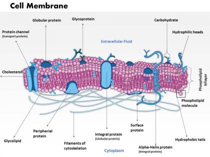
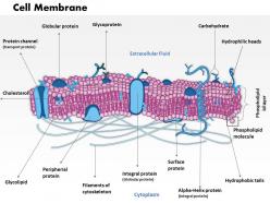
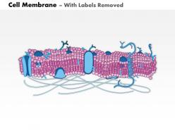
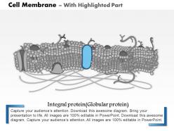
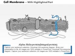
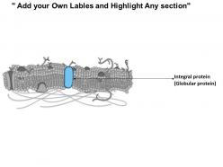
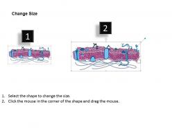
Add cell membrane medical image PowerPoint slide in your presentation. This PPT template has been designed with the graphic of cell membrane and is very valuable to share the information with the medical professionals and students. The structure which includes fluid mosaic model, lipid layer, membrane polarity, membrane structures and cytoskeleton can be easily display with this presentation design. Also, the PowerPoint design can also be helpful in showing the composition as it contains a variety of biological molecules such as lipids, proteins, carbohydrates and phoslipids forming lipid vesicles. The presentation visual perfectly defines function and importance of cell membrane can be shared with the audience with the help of this design. This can be easily modified and you can share your message with it. Furthermore, it is accessible in both standard and widescreen view which allows you to present even among small and large audience. Therefore, download it now and create a visually stunning presentation with this suitable design. Keep your audience totally engrossed. Bait them with our 0714 Cell Membrane Medical Images For PowerPoint.
People who downloaded this PowerPoint presentation also viewed the following :
0714 cell membrane medical images for powerpoint with all 10 slides:
The aim of your thoughts and our 0714 Cell Membrane Medical Images For PowerPoint will always coincide. They have the same aspirations.


FAQs for 0714 cell membrane medical
So the cell membrane is basically your gatekeeper for drug delivery - controls what gets in and what stays out. Some drugs just slip through via passive diffusion, others need transporters or endocytosis to help them along. Size, charge, and how lipophilic the drug is all matter. What's wild is how the membrane composition changes between different cell types, which makes targeting specific tissues such a pain. Oh, and different drugs need totally different approaches. When you're designing delivery systems, figure out the membrane's permeability stuff first - trust me, it'll save you so much trouble later.
Hey! So fluorescent dyes like propidium iodide are perfect for this - they only get through busted membranes. Live cell microscopy is wild because you can literally watch membranes bleb and rupture as it happens. If you need crazy detail, electron microscopy shows all the ultrastructural damage. Calcein-AM works great too for tracking when diseased cells get leaky. MRI contrast agents are useful clinically for tissue work, though that's probably overkill for what you're doing. I'd start with fluorescent viability assays honestly - they're fast, give you solid numbers, and perfect baseline data.
Honestly, super-resolution stuff like STORM and PALM changed everything - you can finally see membrane details that were impossible before. Two-photon microscopy is amazing for going deeper into tissues without killing your samples. Live cell imaging blew my mind when I first tried it; watching membranes move in real time is crazy cool. Oh, and cryo-electron tomography gives you insane detail at basically atomic level. Your core facility probably has some of these options available. I'd definitely check what they've got before planning anything major - could save you tons of headaches later.
So it totally depends on what you're trying to figure out. Fluorescence is perfect for watching live cells - you can literally see proteins moving around in real time, which is pretty cool. If you need crazy detailed structure though, electron microscopy is unbeatable. Your cells will be dead but the images are insane. Confocal lets you slice through membrane layers optically, and super-resolution stuff like STORM gets down to individual protein clusters. I'd probably start with fluorescence to get the basics, then switch to EM if you need those structural details. There's honestly no single "best" option.
Yeah, cell membrane changes are massive in cancer progression - you see it all the time in imaging. Cancer cells basically rewire their membrane composition and make it more fluid, so they can squeeze through spaces they shouldn't be able to. They also jack up receptor expression and mess with signaling right at the membrane. Oh, and it helps them dodge chemo by changing drug absorption - pretty sneaky honestly. When you're looking at images, definitely watch for membrane irregularities and thickness changes. Those are often the first signs something's going malignant.
So basically, the lipids in cell membranes totally control how drugs get through. More cholesterol = stiffer membranes, which makes it harder for fat-loving drugs to slip in. Water-loving drugs have it even worse. The mix of different phospholipids changes how fluid everything is too - that's actually why chemo works better on some cancers than others, which is pretty wild when you think about it. When you're looking at those membrane pics, check for lipid rafts and how thick the membranes are. Those details will tell you where drug delivery might hit roadblocks.
Oh man, there's SO much happening with membrane research right now. Cancer's obviously huge - they're looking at how messed up membranes help tumors spread and resist drugs. Alzheimer's too, since those amyloid plaques just wreck neuronal membranes. Then you've got cardiovascular stuff focusing on endothelial dysfunction. MS and other autoimmune diseases are big since myelin gets destroyed. The imaging tech is insane now - you can literally watch membranes change in real time with confocal microscopy. Honestly feels like every disease has some membrane angle these days, which I guess makes sense when you think about it.
Your cell membranes basically control how everything talks to each other inside your body. If they get too stiff or too floppy, signals can't get through properly - like in diabetes where insulin receptors won't cluster right, or Alzheimer's where cholesterol screws up brain cell communication. It's wild how one tiny change breaks the whole chain. The membrane makeup decides whether hormones and neurotransmitters can actually work. Oh, and those microdomain patterns you see in images? They're probably showing you way more about what's going wrong than you think.
Okay so membrane studies are actually super important for gene therapy - they show you exactly how your delivery vectors get into cells. Different cell types have totally different membrane compositions (which honestly blew my mind when I first learned it). The imaging reveals specific pathways like endocytosis routes and receptor binding that you'd never catch otherwise. You can use this data to pick better targeting ligands and optimize your whole delivery system. Plus it helps predict which tissues will actually take up your genes. Trust me, do the membrane imaging early or you'll hate yourself when vectors fail later.
So AI can really speed things up for membrane analysis - it'll auto-detect structural issues and measure thickness variations way faster than doing it by hand. The pattern recognition is honestly getting ridiculous... these algorithms spot tiny changes in permeability or protein distribution that even experienced radiologists miss sometimes. Plus you can standardize interpretations across different imaging types, which cuts down on that annoying inter-observer variability. I'd say start by adding AI detection tools to whatever workflow you're already using. Just don't get lazy with it - always double-check the results since it works best as backup, not a replacement.
Honestly, membrane imaging is a pain. Those things are crazy thin - like 4-10 nanometers - so regular light microscopy just can't see them properly (hits a wall around 200nm). Electron microscopy helps but the sample prep is brutal. All that fixation and dehydration creates weird artifacts that'll make you question everything you're seeing. Oh, and forget about watching live cells since membranes never stop moving around. I'd probably try cryo-EM first to get the structure down, then maybe throw in some super-resolution fluorescence to catch the dynamics. It's definitely a "use whatever works" situation.
So basically, cell membrane studies are huge for autoimmune research - they show us exactly how immune cells screw up and attack healthy tissue. The imaging is honestly pretty crazy to watch; you can see autoantibodies binding to membrane proteins in real-time. This helps researchers figure out which membrane bits trigger the autoimmune response and how inflammation trashes cellular barriers. Most new autoimmune treatments target membrane proteins because of this imaging work. Pro tip: when you're looking at autoimmune cases, check the membrane integrity markers first. They predict disease progression way better than the usual inflammatory stuff.
So basically, protein distribution shows you if the cell membrane is working right. Different proteins hang out in specific spots to do their jobs - transport stuff, send signals, keep structure intact. When I look at medical images, weird protein patterns usually mean something's wrong before other symptoms even show up. It's kinda like having a crystal ball for cellular health, honestly. You want to check if the key proteins are sitting where they're supposed to be. If they're clumped up weird or missing entirely, that's your red flag for membrane problems.
So basically, cell membrane imaging lets you peek inside and see what healthy membranes actually look like versus the busted ones. Pretty wild stuff honestly - you can spot membrane permeability issues, check protein layouts, see how lipids are organized. All that detail helps you figure out what's broken and needs repair. Then you've got solid data to work with for stem cell treatments or designing better biomaterials. I always thought the communication aspect was fascinating too - how cells talk to each other through these membranes. Anyway, I'd start by checking membrane integrity markers in whatever tissue you're targeting first.
Honestly, STORM and PALM are incredible for membrane imaging - they blow past those old diffraction limits and you can see nanoscale stuff happening live. The fluorescence lifetime imaging is where it gets really cool though, watching proteins interact right at the membrane surface. Cryo-electron tomography has amazing resolution for structural details, but ugh the sample prep is such a hassle. Light sheet microscopy works great for whole tissues if you don't want to mess up the membrane architecture. I'd jump on learning these soon since everything's moving so fast in this field right now.
-
Topic best represented with attractive design.
-
Out of the box and creative design.