


Karyotyping Analysis PPT Graphics ACP





Try Before you Buy Download Free Sample Product
 Impress Your
 Audience
 100%
100% Editable
 Save Hours
 of Time


Elevate your presentations with our Karyotyping Analysis PowerPoint Deck. This professional collection features high-quality graphics, detailed charts, and insightful visuals to effectively communicate complex genetic concepts. Perfect for educators, researchers, and students, it simplifies karyotyping analysis and enhances understanding of chromosomal structures and abnormalities.
People who downloaded this PowerPoint presentation also viewed the following :
Karyotyping Analysis PPT Graphics ACP with all 44 slides:
Use our Karyotyping Analysis PPT Graphics ACP to effectively help you save your valuable time. They are readymade to fit into any presentation structure.












FAQs for Karyotyping Analysis
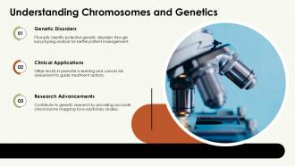
Karyotyping is a laboratory technique that analyzes an individual's complete set of chromosomes to identify structural abnormalities, numerical variations, and genetic disorders. This process enables healthcare institutions, research facilities, and diagnostic laboratories to detect conditions like Down syndrome, Turner syndrome, and chromosomal deletions, ultimately delivering precise genetic counseling and targeted treatment strategies for improved patient outcomes.
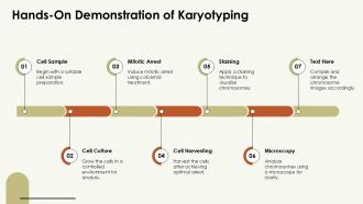
Karyotyping identifies chromosomal abnormalities by analyzing chromosome number, structure, and arrangement through systematic visualization of an individual's complete chromosome set. This diagnostic technique enables medical professionals to detect conditions like Down syndrome, Turner syndrome, and chromosomal deletions, ultimately delivering precise genetic diagnoses and informed treatment planning for patients across healthcare institutions.
Standard karyotyping techniques include G-banding, C-banding, Q-banding, R-banding, and FISH (fluorescence in situ hybridization). These methods enhance chromosome visualization by creating distinctive banding patterns, enabling precise identification of structural abnormalities, and facilitating accurate genetic analysis, with many diagnostic laboratories finding that combining multiple banding approaches ultimately delivers more comprehensive chromosomal assessments and improved clinical outcomes.
Stains and dyes enhance chromosome visualization by creating distinct banding patterns, improving contrast, and highlighting specific structural features that make individual chromosomes identifiable during analysis. Through techniques like Giemsa staining and fluorescent dyes, laboratories can detect chromosomal abnormalities, structural variations, and genetic disorders more accurately, ultimately delivering faster diagnostic services and better patient outcomes in clinical genetics.
Karyotyping in prenatal diagnosis identifies chromosomal abnormalities like Down syndrome, Turner syndrome, and trisomy 18 through analysis of fetal cells obtained via amniocentesis or chorionic villus sampling. This diagnostic approach enables healthcare providers to detect genetic conditions early, counsel families on potential outcomes, and develop comprehensive care plans, ultimately delivering informed decision-making and improved patient management throughout pregnancy.
Karyotyping assists in cancer diagnosis by identifying chromosomal abnormalities like deletions, translocations, and duplications that characterize specific cancer types, enabling precise classification and staging. This chromosomal analysis helps oncologists select targeted therapies, predict treatment responses, and monitor disease progression, with many cancer centers finding that karyotype-guided approaches deliver more personalized treatment plans and improved patient outcomes.
Traditional karyotyping limitations include low resolution preventing detection of small chromosomal changes, inability to identify balanced rearrangements, requirement for dividing cells, labor-intensive processes, and subjective interpretation challenges. While these constraints have historically limited diagnostic precision, many laboratories are increasingly integrating advanced molecular techniques like chromosomal microarrays and next-generation sequencing, ultimately delivering enhanced accuracy and faster diagnostic capabilities for clinical applications.
Digital image analysis enhances karyotyping accuracy by automating chromosome detection, standardizing measurements, and reducing human interpretation errors through advanced algorithms. These technologies streamline analysis by improving contrast enhancement, precise banding pattern recognition, and consistent chromosome classification, with many clinical laboratories finding that automated systems deliver faster diagnostic turnaround times and more reliable genetic assessments.
Metaphase karyotyping examines condensed chromosomes during cell division, providing clear structural visualization but requiring actively dividing cells, while interphase karyotyping analyzes chromosomes in non-dividing cells using fluorescent probes. These complementary approaches enable comprehensive genetic analysis across clinical laboratories, with many diagnostic facilities finding that combining both methods delivers faster results, broader applicability, and enhanced diagnostic accuracy for genetic disorders.
Karyotyping reveals evolutionary relationships by comparing chromosome structures, numbers, and arrangements across species, identifying common ancestral patterns and genetic divergence points. Through chromosomal analysis, researchers trace evolutionary pathways, understand speciation events, and map genetic relationships between organisms, with comparative studies in mammals, plants, and insects ultimately delivering crucial insights into biodiversity and evolutionary mechanisms.
Karyotyping in human genetics raises significant ethical considerations including informed consent complexities, genetic discrimination risks, psychological impacts of abnormal results, privacy concerns, and reproductive decision-making pressures. These challenges present both opportunities for enhanced genetic counseling protocols and considerations around accessibility, with many healthcare institutions finding that comprehensive ethical frameworks ultimately deliver better patient outcomes while maintaining diagnostic accuracy.
Karyotyping varies significantly across species due to differences in chromosome number, size, and structure, with plants often displaying polyploidy and animals typically maintaining diploid sets. While human karyotypes feature 46 chromosomes and mammals show similar patterns, plant species can range from simple diploids to complex polyploids, ultimately enabling researchers to understand evolutionary relationships and genetic diversity across different biological kingdoms.
Technological advancements influencing karyotyping include automated chromosome analysis systems, high-resolution digital imaging, fluorescence in situ hybridization (FISH), comparative genomic hybridization, and AI-powered diagnostic software. These technologies enhance diagnostic accuracy by streamlining chromosome identification, reducing analysis time, and enabling detection of subtle genetic abnormalities, with many genetic laboratories finding significantly improved patient outcomes and faster clinical decision-making.
Karyotyping integrates with fluorescence in situ hybridization (FISH) and SNP arrays through complementary diagnostic workflows that enhance chromosomal analysis precision and resolution. FISH provides targeted visualization of specific genetic sequences on karyotype preparations, while SNP arrays deliver genome-wide copy number analysis, with many clinical laboratories finding that this strategic combination streamlines genetic diagnosis, reduces turnaround times, and ultimately delivers more comprehensive patient assessments.
Non-disjunction events identified through karyotyping reveal critical chromosomal abnormalities that significantly impact genetic counseling, reproductive planning, and clinical decision-making. These findings enable healthcare providers to diagnose conditions like Down syndrome, Turner syndrome, and Klinefelter syndrome, while helping families understand inheritance risks, treatment options, and long-term care strategies, ultimately delivering more personalized and effective patient management.
-
A perfect platform for all your presentation needs. Unlimited products at an affordable price.
-
Design layout is very impressive.