


Clinical Neuroanatomy And Neurophysiology PPT Information ACP





Try Before you Buy Download Free Sample Product
 Impress Your
 Audience
 100%
100% Editable
 Save Hours
 of Time


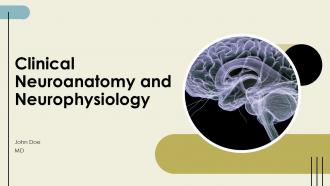
Elevate your understanding of Clinical Neuroanatomy and Neurophysiology with this comprehensive PowerPoint presentation deck. Designed for professionals, it offers in-depth insights, detailed visuals, and essential information to enhance your knowledge and application in clinical settings. Perfect for educators, clinicians, and students in neuroscience.
People who downloaded this PowerPoint presentation also viewed the following :
Clinical Neuroanatomy And Neurophysiology PPT Information ACP with all 40 slides:
Use our Clinical Neuroanatomy And Neurophysiology PPT Information ACP to effectively help you save your valuable time. They are readymade to fit into any presentation structure.










FAQs for Clinical Neuroanatomy And Neurophysiology
The central nervous system comprises the brain and spinal cord, with major structures including the cerebrum, cerebellum, brainstem, and spinal cord segments. These structures enable neurologists and neurosurgeons to localize pathology through clinical examination, with cerebral cortex lesions affecting cognition and motor function, cerebellar damage impairing coordination, and brainstem injuries disrupting vital functions, ultimately delivering precise diagnostic capabilities.
Neuroanatomical pathways provide critical diagnostic frameworks by mapping symptom patterns to specific brain regions, enabling clinicians to localize lesions through deficits in motor, sensory, and cognitive functions. Through pathway analysis, neurologists can differentiate between stroke locations, identify spinal cord compression levels, and distinguish demyelinating diseases, ultimately delivering more precise diagnoses and targeted treatment strategies.
Neuroimaging plays a crucial role in clinical neuroanatomy by enabling real-time visualization of brain structures, identifying pathological changes, and correlating anatomical abnormalities with clinical symptoms. These technologies, including MRI, CT, and PET scans, enhance diagnostic accuracy in neurological disorders, facilitate surgical planning, and improve patient outcomes, with healthcare institutions increasingly finding that advanced imaging streamlines treatment decisions and delivers more precise therapeutic interventions.
Neuroanatomical knowledge guides neurosurgeons in precise localization of pathological tissues, critical structure identification, and optimal surgical approach planning, while minimizing damage to healthy brain regions. Through detailed understanding of neural pathways, vascular networks, and functional areas, surgeons can navigate complex procedures more safely, reduce operative complications, and ultimately deliver better patient outcomes with enhanced recovery rates and preserved neurological function.
Anatomical variations in brain structure significantly impact surgical planning, diagnostic accuracy, and treatment protocols, requiring clinicians to adapt standard approaches for individual patients. These variations influence neurosurgical procedures, neuroimaging interpretation, and therapeutic interventions across specialties like neurology and psychiatry, with many healthcare institutions finding that personalized anatomical assessment ultimately delivers safer procedures and improved patient outcomes.
The peripheral nervous system integrates with the central nervous system through cranial nerves, spinal nerves, and specialized ganglia that relay sensory information inward and motor commands outward. These neural pathways enable seamless communication between brain centers and body tissues, with hospitals and rehabilitation centers finding that understanding these connections enhances diagnostic accuracy, treatment planning, and patient outcomes in neurological care.
Clinical considerations for brain areas involved in motor control include assessing cortical motor regions, basal ganglia function, cerebellar coordination, brainstem reflexes, and spinal cord pathways. These neuroanatomical structures present both diagnostic challenges and therapeutic opportunities, with neurologists finding that systematic evaluation of motor cortex lesions, Parkinson's disease, cerebellar ataxia, and spinal injuries enables targeted rehabilitation strategies, ultimately delivering improved patient outcomes and functional recovery.
Understanding neuroanatomical relationships enables clinicians to predict functional deficits, localize stroke damage, and develop targeted rehabilitation strategies based on affected brain regions and their connections. This knowledge helps medical teams anticipate specific symptoms like speech difficulties or motor impairments, customize therapeutic interventions, and optimize recovery pathways, ultimately delivering more precise treatment plans and improved patient outcomes.
Advances in neuroanatomical research include high-resolution MRI, diffusion tensor imaging, connectome mapping, optogenetics, and single-cell sequencing technologies. These innovations streamline understanding of brain-behavior relationships by revealing precise neural circuits, mapping white matter pathways, and identifying cellular mechanisms underlying cognitive disorders, ultimately delivering enhanced diagnostic accuracy and targeted therapeutic interventions for neuropsychological conditions.
Neuroanatomical changes in Alzheimer's disease include hippocampal atrophy, cortical thinning, ventricular enlargement, and characteristic amyloid plaques and neurofibrillary tangles throughout brain regions. These structural deteriorations correlate directly with cognitive decline progression, with healthcare institutions increasingly leveraging advanced neuroimaging and biomarker technologies to enable earlier diagnosis, personalized treatment planning, and improved patient outcomes.
Developmental neuroanatomy findings influence pediatric neurology by revealing critical periods for brain maturation, identifying vulnerability windows for neurological disorders, and establishing age-appropriate diagnostic criteria. These insights enable pediatric neurologists to differentiate normal developmental variations from pathological conditions, optimize timing for interventions, and predict long-term outcomes, ultimately delivering more precise diagnoses and targeted treatment strategies for developing nervous systems.
Neuroanatomy guides rehabilitation by mapping damaged brain regions to specific functional deficits, enabling targeted therapeutic interventions through motor retraining, cognitive exercises, and sensory stimulation techniques. Through neuroplasticity principles, rehabilitation specialists in hospitals and stroke centers design personalized recovery programs that leverage intact neural pathways, ultimately accelerating functional recovery and improving patient outcomes.
Psychiatric disorders involve neuroanatomical alterations including reduced prefrontal cortex volume, hippocampal atrophy, amygdala hyperactivity, disrupted white matter tracts, and altered neurotransmitter pathways. These structural and functional changes manifest differently across conditions like depression, schizophrenia, and anxiety disorders, with neuroimaging technologies enabling clinicians to identify specific patterns, ultimately enhancing diagnostic accuracy and treatment personalization in psychiatric care.
Neuroanatomy research reveals how pain pathways, neurotransmitter systems, and brain regions like the thalamus, somatosensory cortex, and limbic structures process chronic pain signals. This knowledge enables clinicians to develop targeted interventions including nerve blocks, spinal cord stimulation, and pharmacological treatments, with many pain management centers finding that anatomically-informed approaches deliver more effective relief and improved patient outcomes.
Common misconceptions about neuroanatomy in clinical practice include oversimplifying brain-behavior relationships, assuming strict localization of functions, underestimating neuroplasticity, neglecting individual anatomical variations, and relying on outdated hemisphere dominance theories. These misunderstandings can lead to diagnostic errors and suboptimal treatment plans, with many healthcare institutions finding that comprehensive neuroanatomical education and updated imaging technologies ultimately enhance patient outcomes and clinical decision-making accuracy.
-
“I've always gotten excellent slides from them and the customer service is up to the mark.”
-
“Easy to use. I always wanted to have a quick search for products and SlideTeam has helped me have it.”