


Internal anatomy of liver with branch of portal vein branch of hepatic artey




Try Before you Buy Download Free Sample Product
 Impress Your
 Audience
 100%
100% Editable
 Save Hours
 of Time


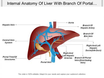
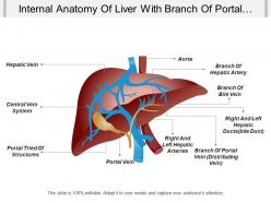
Employ to a greater extent with our internal anatomy of the liver with a branch of portal vein branch of hepatic artery PPT deck and bring folks closer to each other. We have shown the digital interpretation of a human liver and labeled the parts here to give the viewer a clearer picture. Use this design to reflect the health issues in your department and aware your employees regarding the adverse effects. This PPT deck helps the viewer to grasp a clear picture of the layout shown and make it easier for the viewer to understand the data. Design targets output in a standard manner using our, and explain the analysis in your display and get full points. Engage to a greater degree with our Internal Anatomy Of Liver With Branch Of Portal Vein Branch Of Hepatic Artey. Bring folks closer to each other.
People who downloaded this PowerPoint presentation also viewed the following :
Internal anatomy of liver with branch of portal vein branch of hepatic artey with all 5 slides:
Feel extremely fortunate with our Internal Anatomy Of Liver With Branch Of Portal Vein Branch Of Hepatic Artey. The extra bonus will delight you.



FAQs for Internal anatomy of liver with branch of portal vein branch
So the liver's got two main parts - a big right lobe and smaller left one. They're split by this thing called the falciform ligament. Both are packed with these cells called hepatocytes that do the actual work. The right side mostly handles detox and making proteins, while the left focuses on bile and blood sugar stuff. What's wild is how they coordinate everything together - filtering blood, storing vitamins, handling metabolism. If you're dealing with liver issues or just trying to understand how it works, knowing which side does what can really help you figure out what's going wrong.
So the liver's got this pretty smart dual blood supply thing going on. Your hepatic artery pumps in oxygen-rich blood to keep everything alive. Meanwhile, the portal vein brings nutrient-packed blood straight from your intestines - which is honestly genius because the liver gets to process all your food first before it goes anywhere else. It's like having a built-in filter system. Plus there's backup - if one supply gets messed up, the other can kinda pick up some slack. When you're dealing with liver patients, this whole setup totally changes how diseases progress and what surgeons need to think about.
So liver lobules are like these tiny hexagonal factories - maybe a million of them packed in your liver. They're where all the actual work happens. Hepatocytes (liver cells) surround a central vein in each one. Blood flows through these little channels called sinusoids from the edges toward that center. That's how your liver processes nutrients, cleans toxins, makes bile - you know, the usual liver stuff. Pretty cool setup actually. When doctors see liver problems on tests, it usually starts right at this lobular level first. Then spreads outward if things get worse.
Dude, the liver's structure is actually genius when you think about it. Hepatocytes sit in these radiating plates around central veins, so blood flows through sinusoids and hits maximum surface area. Every single liver cell gets direct contact with blood - honestly pretty amazing design. The sinusoids have these fenestrated walls too, which lets big molecules pass through no problem. You've got dual blood supply coming in from both portal vein and hepatic artery, so there's always fresh nutrients and oxygen flowing. When you're studying slides, just focus on how those plates and sinusoids work together. That relationship basically explains everything the liver does metabolically.
So the right lobe is way bigger - like 60% of your whole liver. It does most of the work with glucose storage and making proteins. Left lobe's smaller but still functional. They're split by this thing called the falciform ligament, though surgically it gets more complicated than that looks from outside. Each lobe has its own blood supply and drainage system - that's actually why doctors can remove whole lobes if they have to. Pretty wild when you think about it. Main thing is they're basically two separate units doing the same job, so if you're looking at liver problems, you gotta check both sides since diseases hit them differently sometimes.
So Kupffer cells are these specialized immune cells that hang out in your liver's blood vessels. They're like bouncers, basically - constantly filtering blood coming up from your gut and grabbing onto bacteria, toxins, whatever gross stuff tries to get through. Pretty cool how they're positioned right there to catch things early. They'll also release signals to rally other immune cells when needed. Oh and they clean up dead cell debris too, which sounds gross but someone's gotta do it. The liver's honestly such a workhorse organ when you think about it.
Look for the portal triads first - that's where you'll see hepatic arteries, portal veins, and bile ducts all bundled together. The liver's got this smooth surface with Glisson's capsule covering everything, plus you can spot the falciform ligament splitting the left and right lobes. Those quadrate and caudate lobes are honestly kind of annoying to identify at first. Inside, it's organized into lobules with central veins draining each one. The whole portal triad thing is really what makes sense of how blood flows and bile drains, so start there when you're looking at specimens.
So the liver's spot in your upper right abdomen is actually genius positioning. It sits right under your diaphragm, which means every breath you take massages it and boosts blood flow - nature's pretty smart like that. Your gallbladder tucks underneath for easy bile storage, and being close to your stomach and intestines lets it jump on those nutrients fast. Plus it's right next to major blood vessels for filtering out toxins. Oh, and here's something weird - when patients have liver problems, they'll often complain about right shoulder pain because of that diaphragm connection. Bodies are so interconnected it's wild.
So Glisson's capsule is basically the liver's tough outer shell that keeps everything together when you're moving around or there's pressure changes. It's like a really advanced protective bag around the whole organ. Blood vessels and nerves also run through it to get into the liver tissue - pretty clever design honestly. When the liver gets inflamed or infected, this capsule stretches out and that's what causes that sharp pain in your right upper abdomen. Different from the duller pain you'd get from actual liver tissue problems. Makes sense why it hurts so much when it's irritated.
So basically the liver has this dual venous setup that makes portal hypertension a real pain to deal with. Portal vein carries blood from your gut, spleen, pancreas - then hepatic veins dump everything into the IVC. But when portal flow gets blocked (cirrhosis, clots, whatever), pressure shoots up and blood finds these alternate routes called portosystemic collaterals. That's how you end up with esophageal varices and an enlarged spleen. Honestly, the hepatic venous pressure gradient is probably the most useful measurement you'll get - it tells you actual portal pressure and helps predict who's gonna bleed.
Dude, anatomical variations are everywhere and they'll mess up your surgical planning if you're not careful. Like 45% of cases have weird hepatic artery anatomy, 20% have funky portal vein branching, and bile ducts are anomalous in 15-20%. That "classic" textbook stuff? It's actually pretty rare honestly. Missing these variations can lead to accidental vessel injuries, bleeding, or you might not get complete resections. CT angiography and MRCP are lifesavers for preop planning. Intraoperative ultrasound helps too, especially when things get complicated. Always double-check your imaging beforehand - trust me on this one.
Okay so picture this - hepatocytes arrange themselves in these organized plates with spaces called sinusoids where blood flows through. Between the cells, you've got these tiny channels called bile canaliculi that collect bile right at the cellular level. Pretty cool setup actually. The bile drains from those microscopic channels into bigger and bigger bile ducts. Meanwhile, hepatocytes are basically multitasking - they're filtering blood from the sinusoids while pumping bile into those canaliculi at the same time. It's like they're working both sides simultaneously. Blood processing on one side, bile secretion on the other.
So the liver gets blood from two places - the hepatic artery (oxygenated) and the portal vein (all that stuff from your intestines). Pretty clever setup, right? Everything you eat or drink hits the liver first before going anywhere else in your body. Those sinusoids create tons of surface area for contact, and Kupffer cells basically eat up all the junk and pathogens floating around. But here's the downside - since your liver sees toxins first and at their strongest concentrations, it takes a real beating. That's why so many drugs can mess it up.
So the liver and gallbladder are basically best friends - they're positioned right next to each other and work together constantly. Your liver makes bile all day, then the gallbladder stores it and makes it more concentrated. When you eat something fatty (pizza, anyone?), the gallbladder squeezes and sends that concentrated bile through connecting ducts. It's honestly pretty efficient. The positioning is perfect too - the gallbladder sits in this little groove in the liver. Here's the thing though: if one gets inflamed, the other usually gets cranky too. That's why docs always check both when you've got bile duct issues.
So with cirrhosis, you're basically getting scar tissue that replaces all the healthy liver cells - totally messes up the normal structure. The whole thing becomes hard and bumpy, which is wild to see if you ever look at pathology slides. All that scarring blocks blood flow and causes portal hypertension. Since there's way less functional liver left, it can't make albumin or clotting factors properly anymore. Can't process drugs well either. Here's the tricky part though - these patients can seem okay on the surface while their liver function tests are actually tanking. That's why you've got to keep checking those labs regularly.
-
Nice and innovative design.
-
Awesome use of colors and designs in product templates.