

0514 liver anatomy medical images for powerpoint




Try Before you Buy Download Free Sample Product
 Impress Your
 Audience
 100%
100% Editable
 Save Hours
 of Time


Use our 0514 Liver Anatomy Medical Images For PowerPoint to get the right effect. Influence your audience to follow your lead.
People who downloaded this PowerPoint presentation also viewed the following :
0514 liver anatomy medical images for powerpoint with all 11 slides:
Halve the effort with our 0514 Liver Anatomy Medical Images For PowerPoint. Depend on them to contribute their bit.



FAQs for 0514 liver anatomy medical
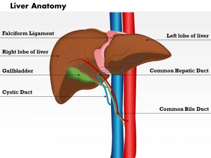
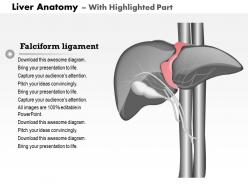
Okay so the liver's got two big lobes - right and left - split by this thing called the falciform ligament. There's also tiny quadrate and caudate lobes but whatever. Hepatocytes are the workhorses doing all the metabolic stuff: processing nutrients, cleaning toxins, making bile. Blood flow is crucial here. Hepatic artery brings oxygen while the portal vein carries nutrient-packed blood from your intestines. Bile ducts drain everything to your gallbladder. Sinusoids let blood flow through while Kupffer cells basically act like bouncers, grabbing toxins and junk. Pro tip: when you're looking at liver cases, follow the vascular patterns first - makes spotting problems so much easier.
So the liver's got this weird dual blood supply thing going on. There's the hepatic artery bringing oxygen-rich blood from your heart - pretty standard stuff. But then you've also got the portal vein, which is kinda cool because it carries all these nutrients straight from your intestines. Most organs don't get that VIP treatment. The portal vein does most of the work though, like 75% of blood flow. Everything drains out through three hepatic veins into the vena cava. Oh, and if you're looking at scans and see portal vein problems? That's bad news since it's basically the liver's main lifeline.
So your liver's basically doing all the heavy lifting for metabolism - breaking down carbs, fats, proteins, plus filtering out toxins. It gets this dual blood supply that's honestly pretty genius. The portal vein dumps nutrient-packed blood straight from your gut, while the hepatic artery brings fresh oxygen. Those liver cells get first access to everything you just ate. The whole thing's structured in these little lobules that maximize surface area for reactions. Kind of like having the world's most efficient processing plant in your abdomen. When metabolic stuff goes wrong, there's usually some liver connection worth looking at.
Think of each lobule like a tiny factory - hepatocytes cluster around a central vein with portal triads at the corners. You get thousands of these working together, which is pretty clever design honestly. The radial setup creates different zones where specific reactions happen in the best spots. Massive surface area too, so every cell can access blood supply and bile drainage easily. I always thought it was wild how this lets the liver juggle so many different jobs at once. That's basically why hepatic function works so well - the whole architecture is built for serious multitasking.
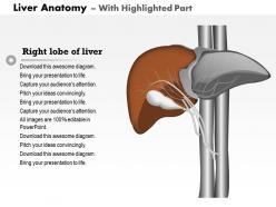
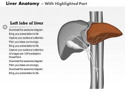
So the right lobe is massive - like 60% of your whole liver - and the left one's smaller and wedge-shaped. Blood supply wise, the right side gets way more flow from the portal vein and hepatic artery. Both lobes do identical work though since hepatocytes are basically the same everywhere. Surgeons hate dealing with the right lobe because it's huge and tucked under your ribs, makes it a pain to reach. Here's the cool part - even though there's such a size difference, they both regenerate equally well. Don't get fooled into thinking bigger automatically means more functionally important.
So hepatic sinusoids are basically your liver's special capillaries - way different from normal ones though. They've got these gaps that let big molecules like proteins pass through easily, which is pretty cool when you think about it. Your liver cells can grab nutrients and toxins straight from the blood this way. Kupffer cells line them too, acting like little cleanup crews. Oh and here's the thing - when these get damaged, your liver performance tanks. That's why doctors always check sinusoid health when they're trying to figure out what's wrong with someone's liver.
So the liver's basically built like the perfect detox machine. Blood flows from these portal areas through tiny channels called sinusoids, then hits the central vein - gives liver cells tons of time to grab toxins. There are these cells called Kupffer cells that just munch on bacteria and junk (honestly pretty cool). Each liver cell sits in single-thick plates so they all touch blood directly for those phase I and II reactions. That's actually why liver damage shows up so fast in blood tests - the whole setup is incredibly efficient but also means there's nowhere for problems to hide.
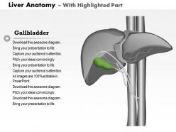
So your liver makes about 500-1000ml of bile every day through hepatocytes. Pretty cool system actually - it flows from tiny bile canaliculi between liver cells, then through bigger intrahepatic ducts, and finally hits the common hepatic duct. From there it either goes straight to your duodenum via the common bile duct or gets stored in the gallbladder through the cystic duct. Here's the thing though - when you're looking at liver problems, figure out if it's a production issue at the cell level or a flow problem in the ducts. That distinction will totally change your diagnostic approach.
So cirrhosis basically destroys your liver's normal structure - instead of smooth, soft tissue, you get these gnarly fibrous bands and nodules everywhere. The liver's trying to heal itself but fails spectacularly. Blood flow gets completely screwed up because scar tissue blocks everything. Function tanks pretty quickly once this starts happening. On imaging, you'll see the liver actually shrinks and looks super irregular - nothing like those perfect anatomy textbook pictures we memorized. Honestly, it's wild how different a cirrhotic liver looks compared to a healthy one.
So the liver's basically built to fix itself, which is wild when you think about it. Hepatocytes can divide super fast when there's damage, and having two blood supplies (hepatic artery plus portal vein) means regenerating tissue gets plenty of nutrients. Those lobular units can work independently while others heal - like having multiple engines. When damage is really bad, stem cells from bile ducts can actually turn into new hepatocytes. I always forget how crazy that is. Bottom line: even nasty liver injuries often heal way better than you'd expect because of all this built-in redundancy.
So basically your liver, pancreas, and gallbladder are like a team. The liver makes bile (basically body soap for fats), then your gallbladder stores it and squirts it out when you eat something greasy. Your pancreas is doing double duty - shooting digestive enzymes into your small intestine while also making insulin that tells your liver what to do with blood sugar. It's actually kind of amazing how they all talk to each other. Oh, and your liver also filters everything you absorb before it hits your bloodstream. That's why when one's acting up, the others usually feel it too.
So basically the liver has this crazy dual blood supply thing going on - it gets blood from both the hepatic artery AND the portal vein. That means it's constantly filtering everything coming from your gut, which is honestly kind of gross when you think about it. But it works! The liver has these specialized immune cells called Kupffer cells lining the blood vessels that just grab onto any nasty stuff floating by. There's also this space called the space of Disse where immune cells can directly touch liver cells. The whole setup processes like 25% of your heart's output while staying on high alert for threats. Pretty wild multitasking.
So the liver sits right under your diaphragm in the upper right side. That's why breathing messes with liver scans - super annoying during procedures honestly. Below it you've got the right kidney, part of the colon, and duodenum all cramped together. The big vein (IVC) runs behind it, and the gallbladder tucks into this little groove on the liver's underside. Here's the thing though - when one organ acts up, the neighbors usually get dragged into the drama too. So if you're looking at liver problems, definitely peek at what's around it.
So the porta hepatis is like the liver's main entrance - everything important goes through there. The hepatic artery brings in oxygenated blood, portal vein carries all the nutrient-packed blood from your digestive system, and the bile duct takes bile out. It's honestly pretty elegant how it's all organized. Without this setup, your liver couldn't do its whole detox thing or handle metabolism properly. Oh and if you're ever looking at liver cases or surgery stuff, this area is crucial - any damage here really messes with the whole organ's function.
So each imaging method shows different liver stuff, which is actually pretty cool. CT scans are great for seeing the overall structure and spotting lesions, especially with contrast. MRI kills it for soft tissue - those T2 images really pop when there's fluid or inflammation. With ultrasound you get real-time blood flow through all the hepatic vessels, which is honestly kind of mesmerizing to watch. I'd say pick CT for general architecture, MRI when you need detailed tissue info, and ultrasound for anything vascular. Really depends what you're trying to figure out.
-
Great product with highly impressive and engaging designs.
-
Easily Editable.