

0514 anatomy of kidney medical images for powerpoint 1




Try Before you Buy Download Free Sample Product
 Impress Your
 Audience
 100%
100% Editable
 Save Hours
 of Time


Get your team on the same bandwidth. Define the spectrum with our 0514 Anatomy Of Kidney Medical Images For PowerPoint 1.
People who downloaded this PowerPoint presentation also viewed the following :
Content of this Powerpoint Presentation
Are you a medical institute or a nephrologist seeking perfect templates to train your students or interns in kidney anatomy? Your search ends here! With these visually appealing, self-explanatory, and well-designed anatomies of kidney PPT templates, you can educate your peers and students on pediatric research on kidney disorders and the diagnosis and management of these kidney diseases. With each part of the kidneys being so clearly showcased, named, and designed, the trainees won't face any obstacles to understanding.
To make the students learn about the kidneys along with the urinary system more deeply and clearly and give them a comprehensive understanding of this biological study, click here to download our professionally designed and colored human kidney urinary system PPT template.
Why go through those extra hassles of designing everything when you can simply take your reliable slide crew's assistance and focus on educating the students with better resources and templates? Dive in!
Template 1: Anatomy of Kidney
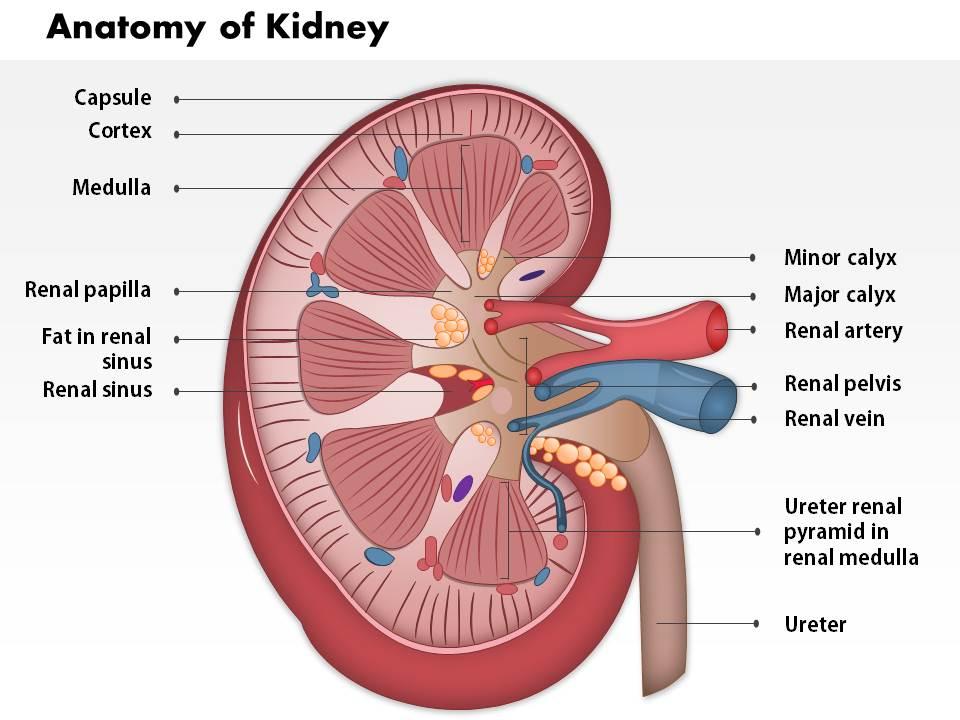
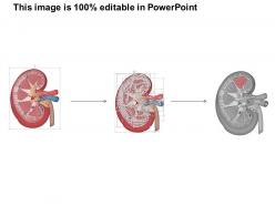
This PPT template presents and simplifies the anatomy of the kidney more clearly than ever before. With the perfect design and visualization, each part of the kidney is accurately labeled and presented to help students understand their functioning. Whether you are a professor or a doctor, this PPT template will simplify your job of presenting a crystal clear understanding of different parts of kidneys, the diseases associated with them, how one could diagnose them, and every essential aspect of anatomy of the kidney to your students and medical professionals.
Template 2: Anatomy Of Kidney - With Highlighted Part
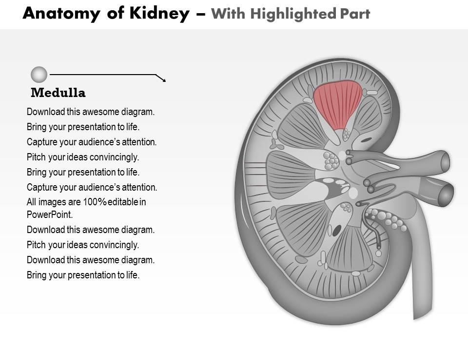
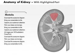
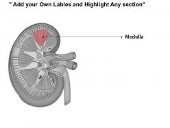
The medulla is one of the important organs in the kidney that controls the urine's concentration. For the students to understand it more precisely, this PPT template will help you impart the importance of medulla in simple words in the slide with an image that particularly highlights that part of the kidney. Simply focus on the medulla and educate your students on it perfectly.
Template 3: Anatomy Of Kidney- With Fat in Renal Sinus
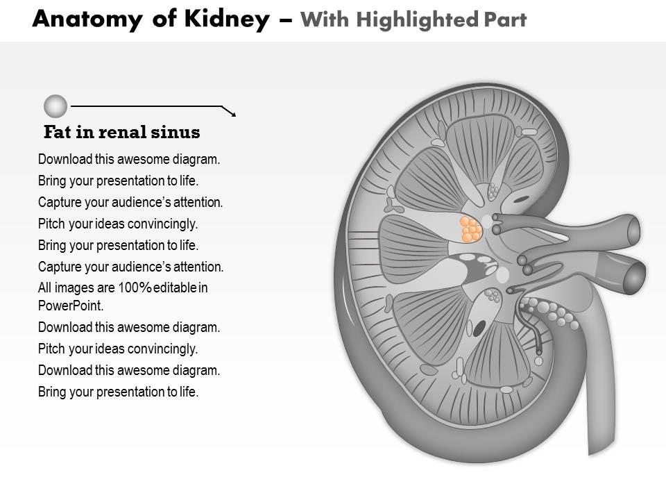
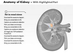
This PPT template will help you to outline the significance of fat in renal sinus with a perfect diagram specially highlighting the fat particles. Educate the interns on how the kidney's renal sinus is a chamber which utilizes fat to protect vital structures like the renal artery, calyces, lymph pathways, and kidney pelvis. The template can also be utilized to present the medical conditions associated with renal sinus such as cysts.
Template 4: Anatomy Of Kidney- Renal Artery
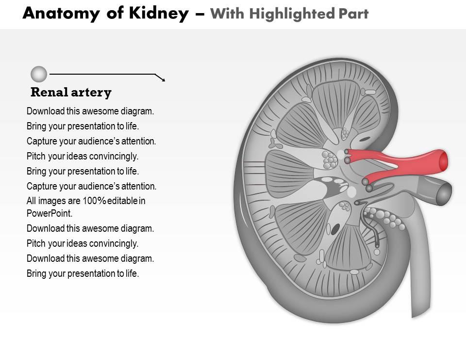
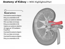
Do you know that every minute, around half a cup of blood flows from the renal arteries through your kidneys? This amazing fact clearly highlights the importance of educating students on the critical role of the human body renal artery in the anatomy of the kidney. This PPT template presents the renal artery and explains how large volumes of blood are transported by them from the kidneys to the aorta, the major artery of the heart.
Template 5: Anatomy Of Kidney - Ureter
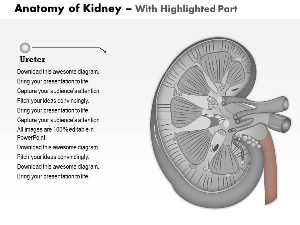
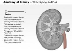
This PPT template will help you showcase one of the important parts of the urinary system- the ureter, through which urine is transported from the kidney to the bladder. With a well-designed diagram slide, you can present the functions and diseases caused by urine flow obstruction, such as stones, infection-related kidney inflammation, etc. The visual design of the PPT focusing on the ureter will simplify your presentation.
The Bottom Line
The kidney is one of the most important organs of our body that helps in blood flow, excretion of urine and waste products and supports the entire urinary system. Thus, it's very important to educate medical students and others on how different parts of the kidney function and how irritation in each can lead to what kinds of disorders. With our professionally curated, neatly designed, and well-labeled PPT templates, you can easily do what is needed. While you prepare your verbal presentations, the Slide Team takes care of your visual presentations.
Ps. Kidney failure can lead to complicated diseases such as diabetes, etc. Click here to get the template that could help you present such crucial issues.
0514 anatomy of kidney medical images for powerpoint 1 with all 11 slides:
Convince them it's not hard with our 0514 Anatomy Of Kidney Medical Images For PowerPoint 1. Your argument becomes easier to believe.


FAQs for 0514 anatomy of kidney medical images
Ok so basically the kidney has three layers - cortex on the outside does the filtering, medulla in the middle concentrates your pee, and then the renal pelvis collects everything before dumping it to your bladder. Each kidney's got like a million nephrons (crazy right?) and they're the actual workhorses doing all the filtering. They're scattered through both the cortex and medulla. Initial blood filtering happens in the cortex, then the medulla tweaks your water and salt levels with those loopy structures. Honestly if you're cramming for an exam, nail down nephron function first - makes everything else click way easier.
So nephrons are like these tiny filters in your kidneys - you've got about a million per kidney which is wild when you think about it. They're constantly filtering your blood and deciding what to keep versus what to toss out. Water, sodium, potassium - they're juggling all that stuff to keep everything balanced. It's honestly pretty cool how they adjust in real time based on what your body needs. Like if you're dehydrated, they'll hang onto more water. Blood pressure regulation happens here too. When doctors run those kidney function tests, they're basically checking if these little guys are still doing their job right.
So the renal capsule is basically this tough outer layer that wraps around your kidney like protective armor. It's surprisingly thin but super strong - keeps the kidney's shape intact and shields it from getting banged up. Also blocks infections from nearby tissues, which is pretty smart if you ask me. When blood flow ramps up, it contains all that internal pressure too. Fun fact: surgeons hit this thing first during kidney operations. Oh, and it's made of fibrous membrane stuff - honestly sounds way more complicated than it probably needs to be, but our bodies love being extra sometimes.
So basically your blood pressure just pushes plasma through these tiny holes in your kidney capillaries. The holes are super small though - water and glucose squeeze through fine, but bigger stuff like proteins get blocked. It's kinda like a coffee filter but way pickier about what passes through. Once it filters through, you've got what becomes your initial urine sitting in this little cup-shaped thing called Bowman's capsule. The whole process is totally passive - no energy required, just pressure doing all the work. Size matters here, and honestly it's pretty cool how selective those little pores are.
So basically the renal pelvis is like your kidney's main collection point - all the urine funnels down there before it heads to your ureter. It's kinda like a storm drain system if that makes sense? Without it working right, you'd get nasty backup and your kidney would be in trouble. The pelvis creates this pressure thing that pushes urine toward your bladder. Oh and if you're ever looking at scans or dealing with blockages, check the pelvis first - that's usually where you'll see it get all swollen up when something's wrong downstream.
So the renal pyramids are where your kidneys do the real work - they've got all the loops of Henle and collecting ducts that concentrate your urine. Filtrate comes down from the cortex and hits this concentration gradient the pyramids create for water reabsorption. That counter-current thing happens here too (which is actually pretty cool). Each pyramid's shaped so it funnels everything toward the renal pelvis through its tip - that's the papilla. Basically it's where your urine gets its final concentration before heading to your bladder. Hope that helps!
So basically your liver is a space hog and pushes the right kidney down about 1-2 cm lower than the left one. Pretty annoying design if you ask me. Both are still the same size though - around 10-12 cm long. The right one also sits closer to your spine compared to the left. It's just another weird body asymmetry thing, like how your heart's off to the left. They're both tucked behind the peritoneum around T12-L3 level. Quick tip for when you're trying to find them: right kidney = lower and more medial. Makes life so much easier during exams.
So basically your kidneys are like really good chemists - they're constantly adjusting your electrolyte levels through these tiny units called nephrons. They'll either reabsorb or dump sodium, potassium, chloride depending on what you need right then. Most of this happens in the distal convoluted tubule and collecting duct (ugh, I know the names are weird). Hormones like aldosterone and ADH help control everything. If you're cramming for a test, definitely focus on how aldosterone works with that sodium-potassium exchange in the collecting duct - that's probably what they'll ask about.
So basically the ureters do all the heavy lifting - they're these tubes that move urine from your kidneys down to your bladder. They're like 10-12 inches long and actually squeeze the urine down using these wave-like contractions (same way your throat pushes food down when you swallow). Gravity alone won't cut it here. The cool part is how they connect to your bladder at this weird angle that creates a natural one-way valve so pee can't flow backward. Which honestly makes sense because that would be... problematic. It's basically an active transport system that's working 24/7.
So your kidneys get about 20% of all the blood your heart pumps out - which is honestly kind of wild when you think about it. Each one has its own direct line from the aorta because they're that important. All that blood flow creates the pressure needed to filter stuff through at around 120 mL/min. When blood flow drops though, everything goes downhill fast. Your filtration rate tanks, you make less urine, and the kidneys start getting damaged. That's why docs always check your BP when they're worried about kidney function - if the plumbing's compromised, the whole system suffers.
So basically the cortex does all the initial heavy lifting - that's where your glomeruli filter blood and grab back all the good stuff your body wants to keep. The medulla takes over after that, concentrating everything through those loops of Henle (weird name, I know) and the collecting ducts to make your final urine. It's like a assembly line kind of thing. Cortex = filtering and cleanup. Medulla = concentration and finishing touches. If something goes wrong with the cortex, you'll have filtration issues. Medulla problems? That's usually concentration troubles.
So basically the kidney gets really good at picking up slack when things go wrong from birth. Remaining tissue just works way harder - nephrons get bigger, they filter more, blood flow shifts around. Honestly it's wild how adaptable they are. Like with renal dysplasia or missing kidneys? The other one can literally double in size and function. Collecting ducts will dilate or reroute flow however they need to. That's why your patients can have normal creatinine for years even with major defects - the compensation is that good. Don't trust basic labs alone for catching this stuff early.
Honestly, I always start with ultrasound - it's fast, cheap, and catches most stones plus any swelling. CT is technically the gold standard, especially CT urography if you want the full picture. MRI works too but mainly when you're worried about radiation or need better soft tissue detail. The thing is, ultrasound finds like 90% of what you're looking for without nuking anyone. CT makes everything super crisp though, I'll give it that. For routine stuff or follow-ups? Definitely ultrasound first. Save CT for when things get weird or you need more answers.
So your kidneys get bossed around by different hormones all day. ADH kicks in when you're dehydrated - it makes your collecting ducts way more permeable so you don't pee out all your water. Aldosterone's job is telling the distal tubules to hoard sodium and ditch potassium. The renin-angiotensin thing is pretty cool actually, it's this whole feedback system that manages blood pressure. PTH handles calcium stuff in the tubules too. None of these mess with the kidney's actual structure - they just flip switches to change how everything works minute by minute.
So the ureters are basically drainage tubes that carry pee from your kidneys down to the bladder using these wave-like contractions. They're about 25-30cm long and have three narrow spots where kidney stones get jammed up constantly - it's honestly annoying how predictable it is. First spot is right where the ureter connects to the kidney, then where it crosses some blood vessels in your pelvis, and finally where it enters the bladder. If they don't work right, urine backs up and you get kidney swelling (hydronephrosis) which can mess up your kidneys permanently. Whenever someone comes in with flank pain, I always check those three bottleneck areas first since that's where problems usually happen.
-
Editable templates with innovative design and color combination.
-
Editable templates with innovative design and color combination.