

0614 human brain side view medical images for powerpoint




Try Before you Buy Download Free Sample Product
 Impress Your
 Audience
 100%
100% Editable
 Save Hours
 of Time


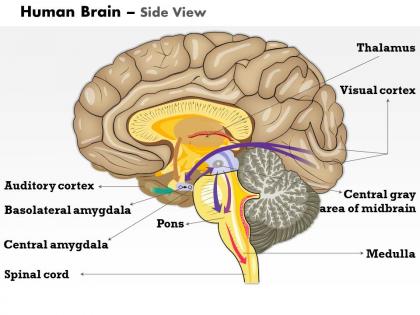
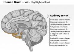
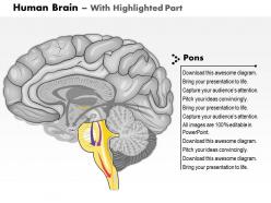
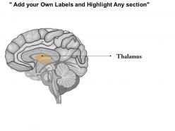
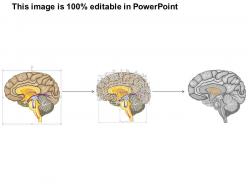
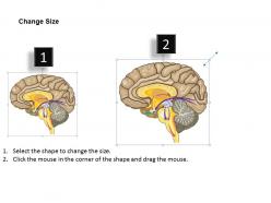
Our 0614 Human Brain Side View Medical Images For PowerPoint add definitive dimensions. They bring to the fore exact descriptions.
People who downloaded this PowerPoint presentation also viewed the following :
0614 human brain side view medical images for powerpoint with all 10 slides:
Add another brilliant dimension with our 0614 Human Brain Side View Medical Images For PowerPoint. You will be a decisive game-changer.
FAQs for 0614 human brain side view medical
So you'll mainly see the cerebral cortex broken into those four lobes - frontal, parietal, temporal, and occipital. The cerebellum's that wrinkly little thing at the bottom back (seriously looks like someone shrunk a whole brain). Then there's the brainstem connecting everything together. Look for the central sulcus first - it separates motor from sensory areas, super helpful landmark. If it's a sagittal cut, you can usually spot the corpus callosum too, that's the bridge between hemispheres. Start by identifying the big divisions, then you can get into the specific gyri patterns later.
Sagittal view is definitely the way to go! You can trace everything from front to back super clearly - prefrontal cortex doing all the executive stuff, then motor/sensory areas, visual cortex at the back. Top or bottom views? Pretty much garbage for understanding function, honestly. Plus you get a great look at the temporal lobe from the side, which makes spotting Broca's and Wernicke's areas way easier. I always forget which is which but whatever - the side view shows both perfectly. Trust me, start with sagittal diagrams when you're explaining this stuff.
So the biggest thing you'll spot is brain size compared to body size. Humans have this huge prefrontal area taking up the front - that's where all our complex thinking happens. Pretty cool actually. Other primates show something similar but way smaller, while most mammals have flatter brain profiles. Cerebellum size changes a lot too, especially in animals with tricky motor skills. Temporal lobes also vary depending on how much auditory processing they need. Focus on those proportional differences when you're comparing, not just raw size. That'll tell you more about how smart different species actually are.
So sagittal brain images are amazing for neurological disease research - you get this clear view from front to back of the brain. Makes spotting atrophy patterns way easier than other angles. The midline structures show up really well too, which is clutch for MS or stroke research. What's cool is you can track how diseases progress over time since everything's laid out in this nice linear way. Plus comparing different patients becomes more straightforward. I mean, lesion locations just pop out more obviously when you're looking at that side profile view.
So you really need good imaging to see brain structures from the side without surgery, obviously. MRI is amazing for soft tissue detail - shows all the different brain regions super clearly in that sagittal view. CT scans work faster and they're better for bones plus spotting bleeding. PET and fMRI can add functional stuff if you want to see what's actually firing. Honestly, picking the right one just depends on what you're looking for. Are you doing research or clinical work? That'll probably determine which type makes the most sense for your project.
I love side views for brain presentations! You can actually see how info flows from the brainstem up through the limbic system to the cortex - that whole "bottom-up" thing becomes super obvious. The corpus callosum looks really dramatic from this angle too, which is cool. Plus it's way easier to show how big different regions are compared to each other. Oh, and definitely try combining it with a top-down view if you can. People seem to "get" brain anatomy much better when they see both perspectives together.
Sagittal brain views are seriously the best for tracking neural connections - way clearer than other angles. You can actually see the corpus callosum bridging both hemispheres and follow white matter tracts between regions. The brainstem-to-cortex pathways are super obvious from this side view. It's kinda wild how you can trace connections from prefrontal areas down to the limbic system. I always check sagittal sections first when I'm looking at connectivity stuff - gives you the whole roadmap before getting lost in detailed imaging. Trust me, start there and work your way into the specifics.
Dude, sagittal views are clutch for trauma cases. The cross-sectional angle shows you exactly how the brain layers got messed up - midline shifts, bleeding, all that stuff jumps out at you. Honestly I always pull these up first now because you catch structural changes so much faster than with other views. Plus when you're talking to families about what happened, they actually get it when you show them the side view. You can literally trace where the injury went and do those before/after comparisons that make everything click. Trust me, start with sagittal slices next time - you won't go back.
Think about it like trying to understand a crumpled up piece of paper by only looking at it from one side - you're missing everything that's folded underneath. The brain has all these layers and pathways weaving through it, but a side view just smooshes it all into a flat image. You can't see how different areas connect or follow those white matter tracts that curve around everywhere. And honestly? Most of the brain's surface is hidden in those wrinkly folds anyway. It's like looking at a 3D maze from just one angle and trying to figure out all the routes. You really need multiple views or 3D imaging to get the full picture.
Side view brain images are perfect for cognitive research - you can spot the prefrontal cortex and temporal structures super clearly. The sagittal view makes it way easier to find Broca's area or trace the default mode network. Way better than getting lost in those axial slices, honestly. You'll see brain activation patterns line up nicely with cognitive tasks since language, memory, and executive areas are right there in view. I always use them as my go-to reference when I'm designing studies or trying to explain findings to people. Makes everything click faster.
Photogrammetry's probably your best bet - Meshroom or RealityCapture can take those side view images and match up points to build depth maps. Structured light scanning works too. Honestly though, most people doing brain modeling use MRI or CT data since you get actual volume info instead of just surface stuff. Neural radiance fields are pretty hot right now if you want to get fancy with it. Just make sure you've got enough overlapping shots or the reconstruction will look like garbage. Oh, and photogrammetry's way easier to get started with than the other options.
Lateral brain images are super helpful for surgical planning - you get this clear picture of how everything sits next to each other. Map out your approach, see what to avoid, figure out the best entry points. Sagittal views especially show depth relationships you can't catch from other angles. It's basically like GPS for brain surgery (scary thought, right?). Short sentences work better sometimes. Anyway, definitely cross-check with your other scans before making final calls. Don't rely on just one view - that's asking for trouble.
So the main ones you need are central sulcus (divides frontal and parietal lobes) and sylvian fissure for the temporal boundary. Corpus callosum shows up nice in sagittal views. Don't miss the brainstem stuff - midbrain, pons, medulla. Cerebellum too if it's in your slice. For functional work, precentral gyrus is your motor area and postcentral is somatosensory. Honestly, I always mess up those two at first glance. These help tons with your coordinate system and ROI analysis. Just make sure you check atlas alignment against these landmarks before diving into anything else.
Honestly, side views are clutch for brain stuff because you can actually see how everything connects. Like, the frontal lobe doing its decision-making thing while your visual cortex processes what you're looking at - and you can literally see the pathways between them. Way better than trying to piece it together from front-on shots. Plus you get that corpus callosum bridge connecting both sides, which is pretty cool. Oh, and it's kind of like watching an assembly line instead of random parts scattered around. I'd start with the big side view in your presentation, then drill down into specifics.
Brain images work so well because they make mental health look medical instead of just "being dramatic." People see the actual anatomy and suddenly realize depression isn't a personality flaw - it's your brain doing brain things. Way more credible than those cheesy stock photos of someone crying by a window, you know? These visuals help normalize talking about ADHD, anxiety, whatever by making it about brain health. Simple messaging works best - just explain which brain parts handle mood and behavior. Honestly, anything that gets people to stop treating mental health like a character weakness is worth trying.
-
Content of slide is easy to understand and edit.
-
Editable templates with innovative design and color combination.