


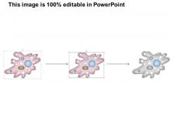
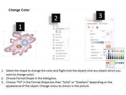
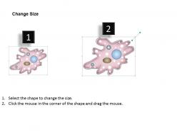
0614 amoeba medical images for powerpoint




Try Before you Buy Download Free Sample Product
 Impress Your
 Audience
 100%
100% Editable
 Save Hours
 of Time


Bounce your thoughts of our 0614 Amoeba Medical Images For PowerPoint They will give you a valuable response.
People who downloaded this PowerPoint presentation also viewed the following :
0614 amoeba medical images for powerpoint with all 11 slides:
Broaden their choices with our 0614 Amoeba Medical Images For PowerPoint. Create excellent avenues of employment.
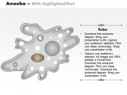
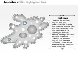
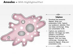
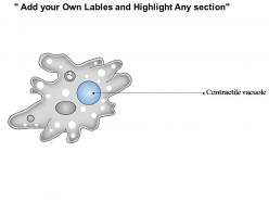

FAQs for 0614 amoeba medical
Medical images produced using amoebae in laboratory research include phase contrast microscopy, differential interference contrast imaging, fluorescence microscopy, time-lapse video microscopy, and electron microscopy techniques. These imaging methods enhance cellular research by revealing amoeba movement patterns, structural changes, and metabolic processes, with many pharmaceutical companies and research institutions finding that such detailed visualization accelerates drug discovery and pathogen studies.
Amoebae contribute to medical imaging development by serving as biological models for studying cellular movement, membrane dynamics, and phagocytosis processes that inform contrast agent design and cellular tracking methods. Through their unique morphological properties, researchers enhance fluorescence microscopy, develop targeted imaging probes, and improve real-time cellular visualization techniques, ultimately delivering more precise diagnostic capabilities for cancer detection and inflammatory disease monitoring.
Amoebic infections present significant diagnostic challenges in medical imaging, requiring specialized MRI, CT, and ultrasound protocols to identify tissue damage, abscess formation, and organ involvement. These parasitic conditions, particularly in liver, brain, and intestinal cases, demand enhanced imaging contrast and resolution techniques, with radiologists increasingly leveraging advanced imaging algorithms to differentiate amoebic lesions from other pathologies, ultimately improving diagnostic accuracy and patient outcomes.
Amoebic motion can be visualized through time-lapse microscopy, fluorescence imaging, phase-contrast microscopy, and advanced digital enhancement techniques that track cellular movement patterns. These imaging methods enable researchers and clinicians to monitor pathogen behavior, assess treatment effectiveness, and study infection progression in real-time, ultimately delivering enhanced diagnostic accuracy and more targeted therapeutic interventions.
Capturing high-resolution amoeba images presents challenges including specimen preparation artifacts, rapid cellular movement during imaging, maintaining viable conditions without fixation damage, optimizing contrast for transparent structures, and managing photodamage from intense illumination. These technical hurdles require specialized microscopy techniques, advanced sample preparation protocols, and careful timing coordination, with many research laboratories finding that combining multiple imaging modalities ultimately delivers clearer morphological data and more reliable diagnostic outcomes.
Different staining techniques significantly enhance amoeba medical image clarity by improving contrast, highlighting cellular structures, and distinguishing between tissue types through targeted dye absorption. Techniques like hematoxylin and eosin create distinct color differentiation, while specialized stains reveal specific organelles and membrane boundaries, ultimately enabling pathologists to achieve more accurate diagnoses and streamline clinical workflows.
Advanced microscopy technologies most effective for studying amoebic interactions include confocal laser scanning microscopy, transmission electron microscopy, fluorescence microscopy, live-cell imaging systems, and scanning electron microscopy. These technologies enable researchers to visualize cellular interactions, track amoebic movement patterns, and monitor tissue invasion processes in real-time, with many medical institutions finding that combining multiple imaging modalities delivers comprehensive insights into pathogenic mechanisms and therapeutic responses.
Artificial intelligence enhances amoeba medical image analysis through automated pattern recognition, machine learning algorithms, advanced image processing techniques, and predictive diagnostic capabilities. These AI technologies streamline identification by detecting cellular anomalies, accelerating diagnosis timelines, and improving accuracy rates, with many healthcare institutions finding that automated analysis delivers faster patient results and enhanced diagnostic precision.
Ethical considerations include informed consent protocols, animal welfare standards in research environments, data privacy protection, equitable access to emerging treatments, and long-term safety evaluation frameworks. While amoebae-based imaging presents minimal direct harm risks, researchers must ensure transparent disclosure of experimental procedures, secure patient data handling, and fair distribution of technological benefits across diverse healthcare systems, ultimately delivering responsible innovation that maintains public trust.
Amoeba medical images help diagnose and understand diseases like amoebic dysentery by revealing parasite morphology, tissue invasion patterns, and infection severity through microscopic examination and advanced imaging techniques. These visual tools enable healthcare professionals to distinguish between different amoeba species, assess intestinal damage, and monitor treatment effectiveness, ultimately delivering faster diagnosis and more targeted therapeutic approaches for patients suffering from parasitic infections.
Amoeba imaging accelerates drug development by enabling real-time observation of cellular responses, membrane interactions, and therapeutic mechanisms at the single-cell level. Through advanced microscopy techniques, pharmaceutical companies can assess drug efficacy, toxicity pathways, and cellular uptake more precisely, ultimately reducing development timelines and costs while enhancing the accuracy of preclinical testing phases.
3D imaging techniques revolutionize amoeba research by providing detailed spatial visualization of cellular structures, pseudopodia formation, membrane dynamics, and organelle distribution in real-time. These advanced methods enable researchers to track movement patterns, analyze structural changes during feeding and reproduction, and study cellular interactions with unprecedented clarity, ultimately delivering deeper insights into amoebic pathogenesis and therapeutic targeting strategies.
**INPUT**: What advancements in microscopy have enhanced the study of amoebae in medical contexts? **OUTPUT**: Advanced microscopy techniques include confocal laser scanning microscopy, electron microscopy, fluorescence imaging, live-cell microscopy, and super-resolution microscopy. These technologies enhance diagnostic accuracy by enabling real-time visualization, detailed cellular structure analysis, and rapid pathogen identification, with clinical laboratories and research institutions finding that improved imaging capabilities streamline diagnosis and treatment protocols. [Word count: 54 words]
Amoebae research contributes to regenerative medicine by providing insights into cellular regeneration, wound healing mechanisms, and tissue repair processes through their remarkable ability to recover from physical damage. These single-celled organisms demonstrate advanced cellular restructuring and membrane repair capabilities, with researchers increasingly finding that amoeba-based studies enhance understanding of stem cell behavior, ultimately delivering breakthroughs in therapeutic applications.
Amoeba imaging's future prospects in personalized medicine include enhanced cellular analysis, improved diagnostic precision, real-time treatment monitoring, and targeted therapeutic development. Through advanced microscopic technologies, healthcare providers can analyze individual cellular behaviors, customize treatment protocols based on specific patient responses, and accelerate drug discovery processes, ultimately delivering more effective treatments and better patient outcomes in an increasingly precision-focused medical landscape.
-
Commendable slides with attractive designs. Extremely pleased with the fact that they are easy to modify. Great work!
-
Out of the box and creative design.