

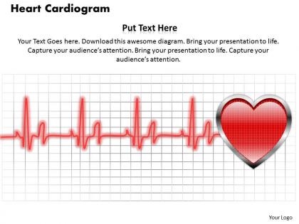
Heart cardiogram powerpoint template slide




Try Before you Buy Download Free Sample Product
 Impress Your
 Audience
 100%
100% Editable
 Save Hours
 of Time


Step out of all boxes with our Heart Cardiogram PowerPoint Template Slide. Reinvent the agendas for tomorrow.
People who downloaded this PowerPoint presentation also viewed the following :
Heart cardiogram powerpoint template slide with all 5 slides:
Meet any demand with our Heart Cardiogram PowerPoint Template Slide. Give greater value to your depth of knowledge.
FAQs for Heart cardiogram
So an EKG is this quick test where they stick electrodes on your chest and limbs to track your heart's electrical activity. Takes maybe 5 minutes tops. The machine spits out this graph with all these wave patterns that doctors read like a roadmap - they can spot irregular rhythms, heart attacks, stuff like that. Honestly, it's pretty cool how those little spikes and dips show whether your heart's pumping blood properly. If you're having chest pain or weird palpitations, they'll probably do this first since it rules out the scary stuff fast.
ECG and EKG are literally the same thing - different abbreviations for electrocardiogram. EKG comes from German "elektrokardiogramm" which is kinda weird but whatever. You'll also hear "cardiac monitor" or "heart rhythm strip" thrown around. Main types are 12-lead ECGs for full heart checks, or continuous monitoring if they need to watch your rhythm ongoing. There's exercise stress ones too, plus those Holter monitors you wear for 24 hours (my aunt had to do that once). If someone orders an "ECG," just ask whether they want a quick one-time reading or the continuous setup.
So basically they stick these electrodes on your skin that pick up the tiny electrical signals your heart makes every time it beats. Pretty crazy that your heart is literally running on electricity, right? The machine amplifies those signals and shows them as those wavy lines you see on the screen or paper. Different electrode spots give doctors different angles to look at what's happening - kinda like taking photos from multiple sides. That way they can catch weird rhythms or if something's damaged. You should totally ask the tech to explain what you're looking at next time!
Okay so basically you're looking for the P wave, QRS complex, and T wave - that's atrial depolarization, ventricular depolarization, and ventricular repolarization. Also check the PR and QT intervals plus the overall rhythm. Each little grid box equals 0.04 seconds which is pretty handy for timing stuff. Not gonna lie, it's intimidating as hell when you first look at one. But seriously, once you practice identifying those basic waves on normal strips, the pattern clicks. Then you can start catching the weird rhythms and arrhythmias. I'd definitely master the basics first though.
So cardiograms show your heart's electrical activity - pretty cool stuff. Getting them regularly lets your doctor catch patterns over time that might mean trouble. It's like... you know how one bad day doesn't define you, but if you're cranky every Tuesday for months, there's probably something going on? Same deal with your heart. You'll catch weird rhythms or medication side effects way earlier. Honestly, the best part is getting your baseline established when you're healthy, then watching for changes down the road. Better to know sooner than later, right?
So basically, EKG patterns show how electricity moves through your heart, and different problems create specific signatures. ST-elevation means active heart attack. Irregular R-R intervals? That's usually a-fib. You'll see inverted T-waves with ischemia or old damage - God, memorizing all these nearly killed me in school. Wide QRS complexes typically mean bundle branch blocks or ventricular stuff. The trick is matching these patterns with what your patient's actually experiencing. Some are pretty obvious once you know what to look for.
So you need 10 electrodes total for a 12-lead. Four go on the limbs - wrists and ankles work, or shoulders/hips if the patient's position is weird. The chest ones are trickier honestly. V1 and V2 go on either side of the sternum at the fourth intercostal space. Then V4 hits the fifth intercostal space at the mid-clavicular line. V3 just goes between V2 and V4 - easy enough. V5 and V6 continue around the left side. Clean skin first or you'll get crappy artifacts. The chest placement definitely takes practice to nail down consistently.
Yeah, patient prep is huge for getting decent cardiogram results. Coffee or running up stairs right before? You'll see a jacked up heart rate that's not even their normal baseline. Anxiety does the same thing - some people freak out just being hooked up to the machine, which is honestly pretty understandable. Poor skin prep is another big one though. Oils, lotions, or even just chest hair can mess with electrode contact and create weird artifacts that look like actual heart problems when they're not. Just have them chill for 5-10 minutes first, skip the caffeine, and get good clean skin contact.
Honestly, AI is crushing it right now with ECG analysis. These algorithms catch arrhythmias and early heart disease signs that doctors might miss - the pattern recognition is insane. Digital machines are way cleaner too, less of that annoying static. But here's what's really wild: wearable monitors give you continuous data instead of those quick 10-second readings. I mean, that's just so much better for actually understanding what's happening. Machine learning keeps getting better at this stuff. If you're trying to boost accuracy, definitely look into AI-assisted software. It's honestly becoming essential for early detection.
Honestly, just be super methodical about it. Rate, rhythm, axis first - then work through intervals, morphology, ST segments. I stick to that order every single time because otherwise you'll miss stuff when you're tired or rushing (learned that the hard way lol). AHA/ESC guidelines are solid to follow. Compare with old ECGs if you have them - sometimes the changes are way more obvious that way. And yeah, don't read in a vacuum - what's the patient actually presenting with? Find whatever sequence works for you and just... never deviate from it. Seriously saves your ass.
So it totally depends on what's going on with your heart. Most people with heart issues go every 6-12 months - I'd say yearly if things are stable. But if you've got heart failure or just had something major happen? Yeah, you'll probably be there every 3-6 months. Honestly feels like you practically live there at first lol. Don't wait for your next appointment though if you're getting new chest pain or weird heartbeats. Just call them and see if you need to come in sooner for an EKG.
So cardiograms are actually pretty amazing for what they can catch. You'll see rhythm problems, signs of old heart attacks, if the heart's enlarged - basically the electrical stuff happening. Arrhythmias and heart blocks show up right away. It's wild how much info you get from those squiggly lines, honestly. They also pick up on things like electrolyte imbalances or if medications are messing with your heart. Obviously it won't catch everything (you'd still need other tests), but for a quick screening? Super reliable first step to figure out who needs more workup.
Yeah, absolutely! Your heart basically gets stronger with exercise, which shows up as better rhythm patterns on EKGs. Cut back on sodium, add more potassium - that helps normalize everything too. Honestly, quitting smoking makes the biggest difference if you smoke. The electrical activity changes are pretty wild within just a few months. Losing weight takes strain off your heart, and you'll actually see improved wave patterns. What's cool is most of these changes show up on cardiograms in like 3-6 months, so it's not forever before you see results. Makes tracking progress way more motivating when you can literally see your heart getting healthier.
Honestly, ECGs are super safe - you're literally just sticking some sticky pads on skin to read electrical signals. No radiation or needles or anything invasive. The only real "risk" is maybe some redness if someone's allergic to the adhesive, but that's pretty rare. Some patients get nervous about it, though I never really understood why since it's way less scary than getting blood drawn. You can tell them it's safer than checking blood pressure. There's basically zero medical risk involved, which is nice for once in healthcare lol.
Oh absolutely - age makes a huge difference with ECGs! Kids have way faster heart rates (like 100-150 for babies is totally fine), plus their PR intervals are shorter and the axis can look weird compared to adults. I used to freak out over pediatric ones until I learned the age ranges. Elderly patients are the opposite - you'll see more baseline conduction issues and atrial changes from just... life, basically. Keep those reference charts close by though. Seriously saves you from calling cardiology at 2am over something that's actually normal for a 75-year-old.
-
Design layout is very impressive.
-
Visually stunning presentation, love the content.