

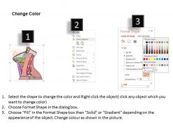
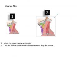
0514 anterior triangle of neck medical images for powerpoint




Try Before you Buy Download Free Sample Product
 Impress Your
 Audience
 100%
100% Editable
 Save Hours
 of Time


Get a good gut feeling with our 0514 Anterior Triangle Of neck Medical Images For PowerPoint. Believe in the fact that your choice is the best.
People who downloaded this PowerPoint presentation also viewed the following :
0514 anterior triangle of neck medical images for powerpoint with all 10 slides:
Financial acumen is your forte. Let your expertise emerge with our 0514 Anterior Triangle Of neck Medical Images For PowerPoint.

FAQs for 0514 anterior triangle of neck medical
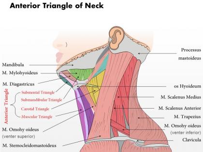
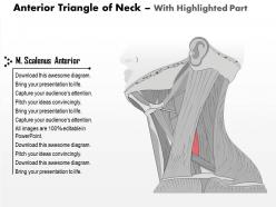
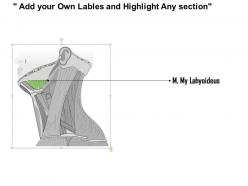
So the anterior triangle has three main borders - sternocleidomastoid muscle on the side, mandible up top, and neck midline medially. Floor's made up of mylohyoid, digastric, and hyoglossus muscles. It's honestly crazy how much stuff gets crammed in there: carotid arteries, jugular veins, cranial nerves, thyroid, lymph nodes. When you're doing dissections or exams, I always start by finding these landmarks first. Makes everything way less confusing. Oh and the visualization part really helps - once you can picture it in your head, the whole region starts making sense.
Yeah, so the anterior triangle is actually pretty variable between people. Neck length and how muscular someone is really changes the whole shape - shorter necks make everything more compressed, longer ones stretch it out. The sternocleidomastoid muscle insertion moves around too, which shifts where that lateral border sits. Honestly, it's kind of wild how different anatomy can be person to person. With heavier patients, you can't even see the triangle boundaries clearly because of all the tissue. My advice? Always feel around for the actual muscle borders instead of just assuming standard proportions. Don't trust your eyes alone - you'll end up missing structures or approaching from the wrong spot.
So the big players in the anterior triangle are your carotid arteries - common, internal, and external - plus the internal jugular vein. The carotid bifurcation is where things get interesting because that's where you'll find the carotid sinus and body. Anterior jugular veins are more superficial, easier to spot. Oh and the facial artery and vein cut through the submandibular part too. Pro tip: always use the carotid pulse as your reference point when you're working in there. Trust me, it'll save you from getting lost in all those deeper structures. Way better than trying to memorize every little landmark.
So the thyroid sits right in that anterior triangle area, which means trouble when things go wrong. Enlarged thyroids will compress your trachea and esophagus - that's where the dysphagia and stridor come from. Those recurrent laryngeal nerves are super close too (surgeons absolutely dread messing with those things), so vocal cord paralysis is always a risk during surgery. You've also got lymph nodes scattered throughout that can pick up thyroid cancer. When you're examining these patients, check for swollen nodes in the whole triangle. Also look for any compression signs - honestly, patients don't always connect their swallowing issues to thyroid problems.
So the anterior triangle has a bunch of cranial nerves doing different jobs. Facial nerve (CN VII) handles your expressions and taste. CN IX does swallowing stuff and throat sensation. Your tongue movement? That's CN XII. The vagus nerve is honestly doing way too much - heart rate, digestion, you name it. People always forget about CN XI but it runs your sternocleidomastoid and trap muscles. When you're checking someone with neck issues, test these systematically. I learned this the hard way - missed nerve dysfunction can totally screw up their swallowing, talking, and facial movements.
So the anterior triangle basically maps out where everything important sits in the neck - your carotid vessels, vagus nerve, lymph nodes, all organized by fascial planes and muscle landmarks. Think of it like a roadmap that keeps you from accidentally cutting something you shouldn't. The subdivisions (submandibular, carotid, muscular) give you the safest way to approach whatever pathology you're dealing with. Honestly, I always sketch these boundaries before any thyroidectomy or neck dissection - helps me spot the danger zones right away. It's probably one of those anatomy concepts that actually matters in real life, you know?
Dude, anterior triangle trauma is seriously scary stuff. You've got the trachea, larynx, and all those major vessels crammed together - any damage there can totally mess up your airway game. Hematomas squeeze everything from the outside while direct hits to the larynx create internal blockages. Intubation becomes a nightmare real quick. Watch for stridor, voice changes, or that crepitus under the skin - those are your red flags. Honestly? I'd rather go straight to a surgical airway than fumble around if there's major swelling. Have your backup ready before you even touch them.
So the anterior triangle is basically lymph node heaven - tons of chains there including deep cervical, submandibular, and submental that drain everything from your head and neck. When you're examining patients, enlarged nodes here are usually your first hint at head/neck cancers. Different spots mean different things too. Like an enlarged jugulodigastric node? Think tonsillar cancer. Submental enlargement might be lip or floor of mouth. I always tell students to palpate systematically from submental down to supraclavicular - don't just randomly poke around hoping you'll find something.
So for anterior triangle imaging, ultrasound is honestly your best bet most of the time. Great for thyroid, parathyroids, lymph nodes - plus it's fast and cheap. CT with contrast works when you need to see deeper stuff or check how vessels relate to other structures. MRI's the gold standard for soft tissue detail, especially with masses or weird lesions you can't figure out. I'd probably start with ultrasound unless there's something specific pushing you toward cross-sectional imaging. Like if you're worried about something deeper that US might miss. Most daily cases though? Ultrasound covers it.
Okay so the anterior triangle is like your cheat sheet for neck pathology. All the important stuff lives there - major vessels, lymph nodes, thyroid, parathyroids, nerves. Honestly the anatomy is pretty dense at first. But here's the thing: once you get those triangular boundaries down, you can actually predict how infections move around and where tumors spread. Different triangles = different symptoms. When I'm looking at neck masses now, I always think in triangles first. Makes the differential way clearer and surgeons actually know what you're talking about when you describe location that way. Trust me, it clicks after a while.
Thyroid stuff is huge in the anterior triangle - nodules, goiter, cancer. Lymph nodes get swollen from infections or cancer spread too. Carotid artery disease pops up there as well. Oh, and submandibular gland stones are annoying but super common - patients always complain about pain when they eat. Parotid issues affect the area even though they're technically more lateral. Honestly, cervical lymph node mets from head/neck cancers are the scary ones you can't miss. When you're examining, just palpate everything systematically. Think inflammatory stuff first, but don't ignore malignancy especially in older folks or smokers.
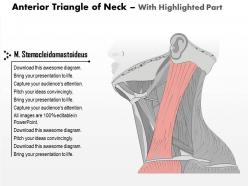
The SCM muscle is basically your cheat sheet for this - it divides the two triangles. Anterior triangle is in front of it, between the midline and jaw. Posterior sits behind the SCM, next to the trapezius. I always found it easier once I got good at feeling for the SCM border. Just trace along it with your finger during exams. Oh, and the anterior triangle has all the good stuff you'll palpate a lot - carotid pulse, thyroid, lymph nodes. The SCM thing honestly becomes automatic after a while.
Honestly, start by finding the carotid sheath, hyoid bone, and thyroid cartilage - they're your best friends for staying oriented. Get through the platysma first, but do it clean. Stay medial to the sternocleidomastoid's anterior border and watch out near the carotid bifurcation because that area gets tricky quick. The recurrent laryngeal nerve sits in the tracheoesophageal groove, so don't go too deep or you'll mess with vocal cords. Palpate everything first and sketch out your game plan mentally - saves you headaches later. Oh, and take your time around the bifurcation, seriously.
So the anterior triangle is where you can actually get to the carotid arteries during an exam. That's your spot for feeling the carotid pulse in the carotid triangle area - honestly pretty straightforward once you know where to look. You'll also listen there for any bruits that might signal stenosis. The sternocleidomastoid muscle makes a nice landmark since it forms the lateral edge. When you're checking someone for possible carotid issues, start there first. Feel the pulse quality, listen for weird sounds, check for anything obviously off. Way easier than trying to remember all the other neck anatomy tbh. Then you can move on to imaging if needed.
Honestly, Netter's Atlas is probably your best starting point - the illustrations are just really solid for getting the basics down. After that, I'd grab Complete Anatomy or Essential Anatomy apps since the 3D models make everything way easier to visualize. AnatomyZone on YouTube has some decent videos too, though some are better than others. If you can get access to cadaveric stuff or ultrasound modules, those are gold because you're seeing actual layers instead of just drawings. Oh, and Grant's Atlas is another good option if you can't find Netter's. Start simple, then work up to the interactive stuff.
-
Topic best represented with attractive design.
-
Unique design & color.