


0714 smallpox virus medical images for powerpoint




Try Before you Buy Download Free Sample Product
 Impress Your
 Audience
 100%
100% Editable
 Save Hours
 of Time


Develop excellent financial foresight with our 0714 Smallpox Virus Medical Images For Powerpoint. Figure out the economy of the future.
People who downloaded this PowerPoint presentation also viewed the following :
0714 smallpox virus medical images for powerpoint with all 9 slides:
Be the best guide with our 0714 Smallpox Virus Medical Images For Powerpoint. Your advice will be clear and compelling.





FAQs for 0714 smallpox virus medical
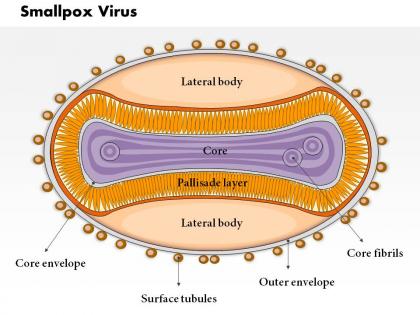
So under electron microscopy, smallpox viruses look like little bricks - seriously, they're pretty distinctive once you spot them. They're huge compared to most viruses (200-400 nm) and have this weird ridged surface that some people say looks like a mulberry, but I've never really seen it. The shape is what gives them away though - rectangular with dense cores and those outer tubules in a crisscross pattern. Honestly, just focus on the size and brick shape first. That'll help you tell them apart from other poxviruses way easier than trying to memorize all the surface details.
Oh man, you can't actually tell the difference just from looking at medical images! Both strains look identical under the microscope - same pustules, same skin patterns, all of it. Kinda wild that something so different clinically looks exactly the same visually. The real difference is how sick people get - major strain is way more deadly and severe, while minor is much milder. But yeah, to know which strain you're dealing with, you need lab work like PCR testing. Images won't cut it - gotta combine clinical symptoms with actual lab results.
Photos are huge for diagnosing smallpox - you need that visual documentation of the lesion pattern. The rash spreads outward from face and hands, which is pretty distinctive. Document everything from multiple angles because smallpox can look surprisingly similar to chickenpox at first. You'll want those images to track how severe it gets and what stage the lesions are in. Honestly, the photos might be your best tool for telling different pox viruses apart. Keep them in clinical records too - epidemiologists will definitely want to see them if there's an outbreak situation.
Smallpox histopathology is actually pretty cool - you can see exactly how the virus tears through tissue. Look for those Guarnieri bodies in epithelial cells, they're like little viral factories going crazy. The progression is wild too, starts with replication then escalates to this massive inflammatory mess that creates the classic skin lesions. Honestly, skin biopsies at different disease stages give you the best view of how systematically this thing destroys normal tissue architecture. The cellular changes are so distinctive once you know what you're looking for.
So cryo-electron microscopy is probably your best bet - it keeps the virus totally natural without all that chemical damage from old fixation methods. Electron microscopy got way better at showing structural details, and atomic force microscopy has crazy good resolution for surface stuff. The fluorescence techniques are pretty cool too since you can actually watch viral components move around during infection in real time. Super-resolution microscopy is solid as well. Honestly, if you're doing poxvirus work, definitely look into the latest cryo-EM protocols. The images they're getting now are just insane.
Yeah, those vaccination scars are usually pretty easy to spot - they look like small, round dents on the upper arm. Having a scar generally means the vaccination worked and you probably have immunity, since it forms from your immune system fighting the live virus. But here's where it gets tricky - you can't really tell someone's current immunity just by looking at a scar in a photo, especially if it's been decades. Some people got the shot but didn't develop long-term protection. Honestly, if you need to know for sure about immunity levels, serological testing is the way to go instead of just eyeballing scars.
So with smallpox images, the lesions go through these stages - macules, papules, vesicles, then pustules. The weird thing is they're all at the same stage everywhere on the body, which honestly makes diagnosis way easier. Chickenpox is totally different - those lesions show up in waves at different stages. Also, smallpox hits the face and arms/legs harder than the chest and back. That's called centrifugal distribution or whatever. If you're looking at suspected cases, those two things - the synchronized development and that distribution pattern - are your biggest clues for telling it apart from other pox viruses.
Medical photos were huge for smallpox eradication - gave officials actual proof of cases and helped everyone diagnose consistently across regions. The WHO's photo archives are wild, you can see surveillance shots from the final cases in Somalia and Bangladesh in the '70s. Field workers used these images to learn the difference between smallpox and chickenpox, which was critical for stopping outbreaks. Countries had to submit visual documentation to verify they'd actually eliminated the disease. Honestly, without that systematic photo tracking, global eradication wouldn't have happened. Definitely worth checking out those archives if you're digging into this topic.
Honestly, the biggest issue is consent - those old smallpox photos were taken way before anyone cared about getting proper permission from patients. So you're basically using someone's worst moments without asking. Those images hit hard too (trust me, seeing them in school was rough). Content warnings are a must. Also think about whether you really need the graphic stuff or if drawings might work instead. Sometimes fewer images actually teach better anyway. Just don't make it sensational, you know? These were real people dealing with horrible illness.
Honestly, infographics are perfect for this stuff. Those medical photos of smallpox are pretty gnarly but they grab people's attention instantly. Then you can layer in the important data - transmission rates, vaccine effectiveness, outbreak timelines. I'd probably show progression photos next to mortality stats by age group, or maybe map out historical outbreaks with vaccination coverage. The visual symptoms paired with hard numbers really drives home why eradication was such a big deal. Start with your most striking image first, then build the public health story around it. People need to see what they're actually dealing with, you know?
Photos are honestly way more useful for diagnosing this stuff. You can see that classic centrifugal pattern - how all the lesions look exactly the same and they're deep in the skin. The uniform pustules on face, arms, hands tell the whole story. Microscopy shows the actual viral particles and inclusion bodies, which is cool I guess, but the rash distribution is so distinctive that photos basically seal the deal. You'd probably want both for proper documentation, but I'd definitely start with good clinical shots. That pattern doesn't lie.
Honestly, digital imaging is a game-changer for smallpox verification. Upload photos to secure platforms and CDC/WHO specialists can analyze them in real-time - we're talking hours instead of days for diagnosis. The image quality now is insane compared to even a few years back. You'll be building a database that helps train both AI and newer clinicians too, which is pretty cool. My advice? Get those secure sharing protocols set up with your regional networks ASAP. Trust me, you don't want to be scrambling to figure this out during an actual outbreak.
Dude, 3D imaging for smallpox research is seriously cool stuff. You can see the virus structure in crazy detail that 2D methods just can't touch. Real-time visualization of how it assembles, replicates, hooks up with host cells - honestly kind of fascinating once you get into it. The detail helps researchers figure out viral mechanisms way better, which obviously matters for vaccines and treatments. I mean, if you're doing any orthopoxvirus work anyway, definitely try incorporating some 3D techniques. The insights about viral architecture will totally change how you think about your experiments.
So basically, those detailed smallpox illustrations are super important for making vaccines work better. They show researchers exactly which surface proteins the virus uses - and that's what our immune system needs to spot and attack. The images help scientists figure out which parts of the virus to actually put in vaccines. Also, they can see how antibodies stick to different viral pieces, which is honestly pretty fascinating stuff. Modern imaging tech captures insane detail now. When you're looking at vaccine studies, check out their methods section - they'll reference these structural images all the time.
Dude, comparative images are seriously your best friend for telling smallpox apart from chickenpox, monkeypox, and all those other blistery diseases. They look so similar at first! But once you see them side-by-side, the patterns jump out. Smallpox lesions are way more uniform - same size, same stage of development. Plus they cluster on the face and extremities instead of mainly on the trunk like chickenpox does. I swear, trying to memorize this stuff from textbooks is useless compared to actually seeing the differences. Keep some reference pics on your phone for when you need them.
-
Excellent design and quick turnaround.
-
Designs have enough space to add content.