


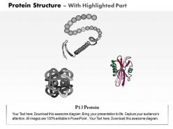
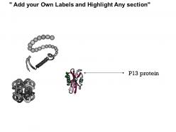
0914 protein structure medical images for powerpoint




Try Before you Buy Download Free Sample Product
 Impress Your
 Audience
 100%
100% Editable
 Save Hours
 of Time


Our 0914 Protein Structure Medical Images For PowerPoint make them gush with adoration. They will behave like crazy fans.
People who downloaded this PowerPoint presentation also viewed the following :
0914 protein structure medical images for powerpoint with all 9 slides:
Get it at a glance with our 0914 Protein Structure Medical Images For PowerPoint. They are absolutely explicit.

FAQs for 0914 protein structure medical
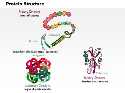
Okay so there are four levels you gotta know. Primary is just the amino acid sequence - like the basic recipe. If that's wrong, you're screwed from the start. Then secondary creates those spiral and sheet patterns (honestly the diagrams make more sense than trying to explain it). Tertiary structure is where it gets interesting - that's the final 3D shape that actually determines what the protein does. Quaternary only matters when multiple protein chains work together as a team. It's kinda like Legos - right pieces, then local patterns, overall shape, and sometimes connecting multiple builds.
So basically, a protein's shape is everything for what it actually does. The 3D structure creates these specific spots where other molecules can bind or reactions happen. Think lock-and-key but way more complex. What's crazy is how even small folding changes can wreck the whole function - that's literally what causes diseases like Alzheimer's when proteins fold wrong. If you're trying to figure out what a protein does, look at its structure first. The shape will pretty much tell you its job. It's honestly the best starting point.
So hydrogen bonds are what keep α-helices and β-sheets from falling apart - they're basically doing all the work. The helix ones are pretty straightforward: carbonyl oxygen bonds with an amide hydrogen that's four spots down the chain. β-sheets are more annoying because the bonds happen between backbone atoms on different strands instead. I always forget that detail tbh. But yeah, without these bonds your protein would just be some random wobbly chain with zero structure. Those dotted lines in protein diagrams? That's the hydrogen bonds holding everything together.
Dude, pH and temperature changes will absolutely wreck protein folding. Basically what happens is the weak bonds holding everything together just fall apart. Change the pH and you're messing with how charged the amino acids are - goodbye ionic bonds. Heat does the same thing but different - like when you cook an egg, right? The proteins literally unfold from all that molecular chaos. Cold temps slow everything down instead. Most proteins are pretty picky about their conditions honestly. I learned this the hard way in undergrad when half my samples turned into useless goop. Keep your buffers steady and don't let temperatures swing around if you want functional protein.
So there's basically three main ways to figure out protein structures. X-ray crystallography gives you insane detail, but honestly growing crystals is such a nightmare - like, sometimes it just won't work no matter what you do. NMR's solid for smaller proteins and you can see how they actually move around in solution. Cryo-EM is huge now for big protein complexes and membrane stuff. Oh, and definitely check the PDB database first before you start anything - might save you months of work if someone already solved it. AlphaFold's predictions are getting scary good too these days.
So chaperone proteins are basically molecular babysitters for newly made proteins. They don't tell the protein what shape to be - that's all in the amino acid sequence. But they stop proteins from clumping together and give them a safe space to fold properly. Heat shock proteins like Hsp70 are the big players you'll hear about constantly. GroEL too, though that one's more in bacteria. Think quality control but for protein folding. If you're doing protein work and getting inclusion bodies instead of nice folded protein, chaperones might be your issue.
So basically proteins have these reusable chunks called motifs and domains that show up everywhere. Motifs are tiny - like binding spots. Domains are way bigger and can actually work on their own. Evolution just keeps copying what works, which is honestly pretty smart when you think about it. That's why you'll find the same domain doing identical jobs in totally different proteins. When I'm looking at a new protein sequence, spotting these familiar pieces instantly gives away what it probably does and how it evolved. It's like recognizing LEGO blocks in different builds.
Yeah, so mutations can really screw things up depending on where they happen. Some are totally harmless and won't even touch the protein structure. But if you hit critical spots - like disulfide bonds or the hydrophobic core - you're basically looking at a folded disaster. The protein might misfold or just stop working entirely. Honestly, predicting which ones will be bad is super tricky. I'd probably run some computational stuff first to get a rough idea, then actually test it in the lab if you need to know for sure. Secondary structure regions are usually pretty sensitive too.
So basically, proteins in your brain start folding wrong and clump up into toxic blobs. Alzheimer's gets those amyloid plaques and tau tangles, while Parkinson's has alpha-synuclein forming these things called Lewy bodies. Pretty crazy that tiny proteins can mess you up so badly, right? These clumps kill your neurons over time - that's why people lose memory, can't move properly, thinking gets fuzzy. Scientists think if we can figure out how to stop proteins from misfolding, we might actually beat these diseases. Or at least slow them down significantly.
So protein denaturation happens when a protein loses its 3D shape and unfolds - basically becomes useless. Heat does it, pH changes, chemicals, even physical stress. You know how egg whites go from clear to white when cooking? That's it right there. The amino acid sequence doesn't change, but all those folded structures get messed up. Shape is everything for proteins - without it, enzymes can't work, structural stuff falls apart. I learned this the hard way in biochem lab when I let samples get warm. Keep your proteins cold and at the right pH if you're working with them!
Okay so think of it like this - enzymes are basically super picky locks that only work with specific keys (substrates). The 3D shape has to match perfectly, plus the charges and chemical stuff all need to line up just right. Honestly, it's crazy how exact everything has to be. One tiny mutation or wrong pH? Boom, your enzyme's useless because the binding site gets warped. I learned this the hard way in lab once - spent hours wondering why my assay wasn't working when it was just the buffer conditions screwing up the protein structure. Always check that first!
Dude, AlphaFold and RoseTTAFold are absolutely crushing it right now - these AI tools predict protein structures crazy accurately. You can literally design brand new proteins from scratch for whatever function you need. Custom enzymes, new therapeutics, the whole deal. CRISPR's been huge for editing proteins in living systems too, obviously. Honestly the computational design platforms are insane for speeding things up if you're working on protein stuff. Oh and directed evolution got way more sophisticated - researchers can optimize proteins for specific traits super fast now. I swear this field moves faster than I can keep up with sometimes, but that's probably a good problem to have.
So quaternary structure is when multiple protein chains come together to make one big functional unit. Hemoglobin's the go-to example - it's got four separate subunits that work as a team. What's really neat is how these subunits can "talk" to each other through allosteric effects. Like, something binds to one subunit and suddenly changes how another one behaves. I always forget this exists until I'm staring at some complex protein structure. When you're dealing with multi-subunit proteins, think about how the arrangement affects what the whole thing actually does.
So post-translational modifications are like your protein's final touch-ups after translation. They totally determine the actual structure and function. Phosphorylation adds negative charges that shift conformations around. Glycosylation helps with folding and keeps things stable. Then you've got ubiquitination - that one's wild because it can completely change where proteins hang out or just mark them for death. Don't sleep on acetylation either, especially with histones. The cool part? Most of these are reversible, so protein structure isn't locked in forever. Honestly, whenever I'm stuck on protein analysis, checking documented PTMs usually explains why my results look nothing like the predicted structure.
Dude, structural variations can make or break your therapeutic protein development. Tiny changes in folding or glycosylation? They'll completely mess with binding affinity and stability. I've seen proteins that look identical on paper act totally different in patients - it's crazy how sensitive these molecules are. You've gotta use mass spec, NMR, cryo-EM throughout development to catch variants early. Don't wait until later stages because by then you're screwed if something's off. Even small aggregation differences can trigger immune responses you definitely don't want.
-
Designs have enough space to add content.
-
Great product with highly impressive and engaging designs.