


0614 whole cell recording medical images for powerpoint


Try Before you Buy Download Free Sample Product
 Impress Your
 Audience
Editable
of Time


The color combos of our 0614 Whole Cell Recording Medical Images For PowerPoint have an advantage. They bring in a carnival effect.
People who downloaded this PowerPoint presentation also viewed the following :
0614 whole cell recording medical images for powerpoint with all 9 slides:
Get a glimpse of the economy of the future with our 0614 Whole Cell Recording Medical Images For PowerPoint. You will acquire financial foresight.
-
0614 whole cell recording medical images for powerpoint
-
0614 whole cell recording medical images for powerpoint
-
0614 whole cell recording medical images for powerpoint
-
0614 whole cell recording medical images for powerpoint
-
0614 whole cell recording medical images for powerpoint
-
0614 whole cell recording medical images for powerpoint
-

0614 whole cell recording medical images for powerpoint
-
0614 whole cell recording medical images for powerpoint
-
0614 whole cell recording medical images for powerpoint
FAQs for 0614 whole cell recording medical
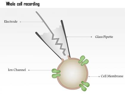
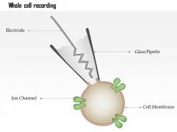
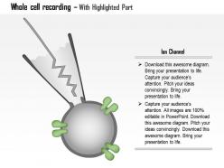
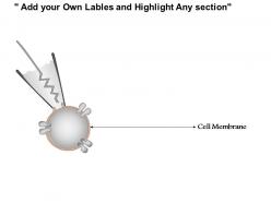
Whole cell recording operates by establishing direct electrical access to a cell's interior through a glass micropipette that forms a seal with the cell membrane, enabling measurement of ionic currents across the entire membrane. This technique delivers comprehensive data on cellular electrical activity, membrane properties, and ion channel function, with researchers in neuroscience, cardiology, and pharmacology finding it essential for understanding cellular mechanisms and drug effects.
Whole cell recording provides direct access to intracellular voltage and current measurements by forming a tight seal with the cell membrane, while extracellular techniques capture signals from outside cells. This approach enables researchers to measure precise membrane potentials, ion channel activities, and synaptic responses with significantly higher resolution and control, ultimately delivering more detailed cellular insights for pharmaceutical development and neurological research applications.
Essential preparations include selecting healthy cells with intact membranes, preparing appropriate intracellular and extracellular solutions, calibrating micropipettes with proper resistance, and establishing stable temperature and pH conditions. These methodical preparations streamline experimental protocols by ensuring consistent seal formation, minimizing noise interference, and maintaining cellular viability throughout recordings, ultimately delivering reliable electrophysiological data and reproducible results across research applications.
Gigohm seal formation requires clean micropipettes with 1-5 megohm resistance, gentle positive pressure during cell approach, and gradual suction application once contact is established. Through careful pressure manipulation and optimal salt solutions, researchers achieve tight membrane-pipette junctions exceeding one gigohm resistance, with many electrophysiology labs finding that patient technique and vibration-free environments ultimately deliver consistent, high-quality recordings essential for accurate cellular measurements.
The patch clamp amplifier controls membrane voltage, measures ionic currents, and amplifies tiny electrical signals during whole cell recording, enabling precise cellular electrophysiology measurements. This sophisticated equipment delivers high-resolution current detection, voltage control capabilities, and signal conditioning, with research laboratories finding that modern amplifiers enhance data quality, streamline experimental workflows, and ultimately accelerate breakthrough discoveries in cellular function analysis.
Whole cell recording enables detailed ion channel analysis by measuring total ionic currents across entire cell membranes, providing comprehensive data on channel kinetics, voltage dependencies, and pharmacological responses. Through voltage-clamp techniques, researchers can isolate specific channel types, quantify current amplitudes and gating properties, ultimately delivering precise insights into cellular excitability and membrane function across diverse physiological conditions.
Common experimental challenges in whole cell recordings include seal formation difficulties, membrane rupture during breakthrough, current rundown over time, space clamp limitations, and dialysis of intracellular components. These technical hurdles affect data quality by compromising electrical access, altering cellular physiology, and limiting recording duration, with many electrophysiology labs finding that systematic troubleshooting protocols significantly improve experimental success rates and data reliability.
Internal solution composition critically affects whole cell recordings by determining ionic gradients, membrane potential stability, and cellular signaling pathways, with different buffers, calcium concentrations, and ATP levels directly influencing recording quality. Researchers must carefully balance solutions for specific applications, as neurobiologists use high-potassium solutions for action potential studies while cardiac researchers employ calcium-buffered solutions, ultimately delivering more accurate physiological measurements and reliable experimental outcomes.
Whole cell recording measures membrane potential, current flow, capacitance, and resistance across cellular membranes, along with intracellular calcium levels and synaptic transmission patterns. These techniques enable researchers in pharmaceutical development, neuroscience labs, and cardiac research facilities to quantify ion channel activity, drug responses, and cellular excitability, ultimately delivering precise data for therapeutic development and disease understanding.
Whole cell recording offers direct access to intracellular neuronal activity, enabling precise measurement of membrane potentials, ionic currents, and synaptic responses with exceptional temporal resolution. This technique streamlines research by allowing simultaneous recording and manipulation of cellular components, with neuroscience laboratories finding that it delivers comprehensive data on neuronal function, synaptic plasticity, and cellular mechanisms, ultimately enhancing understanding of neural circuits.
Whole cell recording combines with fluorescent imaging through simultaneous patch-clamp electrophysiology and optical monitoring, enabling researchers to correlate electrical activity with cellular processes like calcium dynamics, protein interactions, and membrane trafficking. This strategic combination delivers enhanced data resolution, real-time visualization of cellular responses, and comprehensive analysis capabilities, with neuroscience and pharmaceutical research laboratories finding that integrated approaches accelerate drug discovery and cellular mechanism understanding.
Neurons, cardiomyocytes, pancreatic beta cells, immune cells, and smooth muscle cells are most commonly studied using whole cell recordings. These cell types enable researchers to investigate electrical properties, ion channel function, and cellular responses across diverse physiological systems, with many laboratories finding that this technique delivers precise measurements of membrane currents, ultimately advancing drug discovery and therapeutic development.
Whole cell recording limitations include membrane disruption during patch formation, altered intracellular composition through dialysis, limited recording duration due to seal degradation, and potential artifacts from series resistance. While it provides excellent voltage control and direct intracellular access, many researchers find that cell-attached or perforated patch methods preserve more physiological conditions, ultimately delivering less invasive measurements with maintained cellular integrity for longer experimental periods.
Whole cell recording has evolved through automated patch-clamp systems, high-throughput platforms, improved amplifier sensitivity, and advanced data acquisition software. These technological enhancements streamline experimental workflows by enabling simultaneous multi-cell recordings, reducing manual variability, and accelerating data analysis, with pharmaceutical companies and research institutions finding that modern systems deliver faster drug screening and more precise cellular characterization.
Whole cell recording safety precautions include proper electrical grounding, using low-voltage equipment, maintaining sterile conditions, wearing protective equipment, and ensuring proper ventilation. These protocols streamline laboratory operations by preventing contamination, electrical hazards, and exposure risks, with many research institutions finding that comprehensive safety measures ultimately enhance experimental reliability and researcher wellbeing while minimizing costly protocol disruptions.
-
Unique research projects to present in meeting.
-
Professional and unique presentations.