


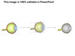
0514 human eye anatomy medical images for powerpoint




Try Before you Buy Download Free Sample Product
 Impress Your
 Audience
 100%
100% Editable
 Save Hours
 of Time


Our 0514 Human Eye Anatomy Medical Images For PowerPoint will give you a different look. Exhibitan air of confidence.
People who downloaded this PowerPoint presentation also viewed the following :
0514 human eye anatomy medical images for powerpoint with all 11 slides:
Lay out the net with our 0514 Human Eye Anatomy Medical Images For PowerPoint. Get the catch of the day with our 0514 Human Eye Anatomy Medical Images For PowerPoint.


FAQs for 0514 human eye anatomy medical
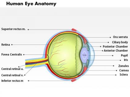
The main components of the human eye include the cornea, lens, retina, iris, pupil, and optic nerve, each serving critical visual functions. The cornea and lens focus light, the retina converts light to electrical signals, the iris controls light entry, and the optic nerve transmits visual information to the brain, ultimately delivering comprehensive sight capabilities.
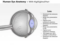
The cornea is a transparent, dome-shaped outer layer providing fixed focusing power, while the lens is a flexible, biconvex structure that changes shape for variable focusing. The cornea contributes approximately 65-75% of the eye's total refractive power through its curved surface, whereas the lens fine-tunes focus by adjusting thickness, enabling clear vision at varying distances.
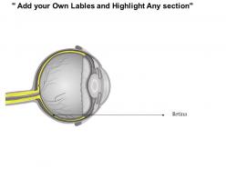
Photoreceptors are specialized cells in the retina that convert light into electrical signals, comprising rods for low-light vision and cones for color detection and sharp detail. These cells contain light-sensitive proteins and are strategically distributed across the retinal surface, with cones concentrated in the fovea for maximum visual acuity, ultimately enabling comprehensive sight processing.
Light travels through the eye by first entering the cornea, passing through the aqueous humor to the pupil, then through the lens and vitreous humor before reaching the retina. This optical pathway enables precise focusing and image formation, with the cornea and lens working together to refract light accurately, ultimately delivering clear visual information that enhances our ability to navigate and interact effectively with our environment.
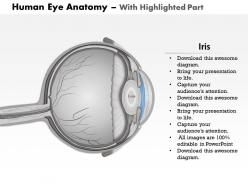
The pupil serves as the eye's adjustable aperture, controlling light entry through automatic dilation and constriction via the iris muscles responding to brightness levels, emotional states, and focusing demands. This dynamic regulation enables optimal vision across varying conditions, from bright outdoor environments to dim indoor spaces, while protecting the sensitive retina from damage and ensuring consistent visual clarity for daily activities.
The six extraocular muscles control eye movement through coordinated contractions, enabling horizontal, vertical, and rotational tracking essential for visual focus and depth perception. These muscles work in synchronized pairs to facilitate smooth pursuit movements, saccadic jumps, and convergence, ultimately delivering precise visual tracking capabilities that enhance spatial awareness and operational efficiency in visually-demanding environments.
Rods and cones differ significantly in function and distribution, with rods specializing in low-light vision and peripheral detection while cones handle color perception and sharp central vision. Rods dominate the retina's outer regions with approximately 120 million cells, whereas cones concentrate in the fovea with around 6 million cells, ultimately delivering complementary visual capabilities for comprehensive sight.
The optic nerve transmits visual information by converting light signals captured by retinal photoreceptors into electrical impulses, which travel through approximately one million nerve fibers to the brain's visual cortex. This neural pathway enables rapid processing of visual data, color recognition, and depth perception, ultimately delivering the comprehensive visual experiences that guide human navigation, decision-making, and daily activities.
Common anatomical variations include iris color differences, epicanthic fold presence, palpebral fissure width, corneal diameter variations, and refractive error prevalence across populations. These variations influence optical characteristics, visual field ranges, and susceptibility to specific conditions like glaucoma or myopia, with many ophthalmologists finding that understanding population-specific traits enhances diagnostic accuracy and treatment personalization.
The tear film protects eye anatomy by lubricating the corneal surface, washing away debris and pathogens, and providing essential nutrients and oxygen to surrounding tissues. This three-layered system maintains optical clarity, prevents infections, and supports cellular health across the cornea and conjunctiva, ultimately ensuring optimal visual function and long-term eye health.
The vitreous humor maintains the eye's spherical shape, provides structural support to the retina, and transmits light from the lens to the retina. This gel-like substance occupies about 80% of the eye's volume, cushioning internal structures against impact while ensuring optimal light transmission, ultimately enabling clear vision and protecting delicate retinal tissues from mechanical damage.
Age-related changes significantly affect eye anatomy through lens hardening, retinal thinning, reduced pupil responsiveness, and decreased tear production, leading to presbyopia, reduced night vision, and dry eyes. These anatomical modifications impact visual acuity and focusing ability, with many individuals finding that regular eye examinations and corrective measures help maintain functional vision and quality of life.
The macula and fovea are critical for sharp, detailed central vision, with the fovea containing the highest concentration of cone cells responsible for color discrimination and fine visual detail. These structures enable precise tasks like reading, driving, and facial recognition, with damage to either region significantly impacting visual acuity and quality of life, making their health essential for maintaining optimal vision.
Common eye diseases impact anatomical structures through inflammation, pressure changes, tissue degeneration, and structural damage to specific components like the retina, cornea, and optic nerve. Conditions such as glaucoma increase intraocular pressure affecting the optic nerve, while macular degeneration deteriorates retinal tissues, and cataracts cloud the lens, ultimately compromising visual function and requiring targeted medical interventions.
The anterior segment contains the cornea, iris, lens, and aqueous humor, while the posterior segment houses the vitreous humor, retina, and optic nerve. These anatomical divisions enable different clinical approaches and surgical techniques, with ophthalmologists finding that anterior procedures like cataract surgery and posterior treatments like retinal repairs require specialized equipment and expertise, ultimately delivering comprehensive eye care across multiple conditions.
-
Helpful product design for delivering presentation.
-
Informative presentations that are easily editable.